Chapter 33 Vascular imaging of the head and neck
The vasculature of the head and neck is now most commonly imaged using techniques other than conventional catheter angiography. The use of computed tomography (CT), magnetic resonance imaging (MRI) and ultrasound techniques allows these vessels to be visualised in a less invasive manner. Catheter studies are still performed in specialist centres, especially as part of endovascular treatment. In this chapter consideration will be given to the use of all these, with some examples of the common indications for imaging.
Common indications for vascular examination
Cerebral aneurysm
This is the most common indication for cerebral angiography. Aneurysmal rupture occurs in 6–12 per 100 000 population and the presence of asymptomatic aneurysms is thought to be in the region of 2% of the population.1 A ruptured aneurysm presents the commonest cause of subarachnoid haemorrhage (SAH) in adults. The most common type is the saccular or berry aneurysm. Typically, defects develop due to the pressure of systolic waves causing herniation of the vessel wall.2
The average age of presentation is 40 years. Below this age presentation is more common in men than in women, but this reverses from 40 years upwards.1 Over 90% of saccular aneurysms occur in the anterior circulation at branch points in the carotid supply,3,4 the remaining 10% being in the posterior circulation.1 In the anterior circulation approximately 25% are located in the middle cerebral artery distribution, 35% around the anterior cerebral artery and 30% associated with the internal carotid artery.1,4,5 Cerebral aneurysms can range from 1–2 mm to 1–2 cm,5 with the risk of bleeding generally increasing with size.1,4,6
The clinical presentation of rupture leading to SAH includes:
Arteriovenous malformation (AVM)
AVMs are the second most common cause of SAH in adults. They result from developmental abnormalities of arterial and venous vessels leading to the formation of fragile vascular walls2 as well as the lack of development of a capillary bed.1 Typically they are made up of three parts: a core of dysplastic vessels known as a nidus, arterial feeding vessels and draining veins.10 The associated vessels are hypertrophic and hyperplastic. AVMs are typically 3–4 cm in diameter2 and are usually symptomatic by the age of 40 years.1
Points of note
• A SAH occurs when blood escapes into the subarachnoid space, which is the space between the arachnoid and the pia mater. In this space it mixes with the cerebrospinal fluid
• A subdural haemorrhage is the presence of blood between the dura and the arachnoid mater
• An extradural haemorrhage is a bleed outside the dura mater, between the dura mater and the bony skull
• SAH may arise from a ruptured intracranial aneurysm, a bleeding AVM, or infrequently as a result of trauma. In contrast, subdural and extradural haemorrhages usually occur as a result of trauma
Stroke
The World Health Organization has estimated that, worldwide, there were 5.7 million deaths from stroke in 2005, equivalent to 9.9% of all deaths.11 A first or recurrent stroke is experienced by 110 000 people in England each year,12 and stroke accounts for 11% of all deaths in England and Wales.13 In England there are more than 900 000 people living with the effects of stroke.13
Stroke may be divided into two main categories:
Ischaemic stroke
This type may lead to regional infarction or to small isolated areas known as lacunar infarcts. It occurs as a result of a regional lack of blood supply to the brain and can be due to occlusion of an artery by any mechanism, such as thrombosis, emboli or dissection. Thrombus may form intracranially or more commonly at the region of the carotid bifurcation, a common site for atheromatous disease and from where distal emboli frequently occur.
Haemodynamic ischaemic stroke occurs following:
• a reduction in perfusion for any reason
• dissection of the vessels in the neck particularly following trauma.
Vasospasm, a common complication of SAH, may also lead to ischaemic stroke. This process can also be a complication of the use of some recreational drugs.
Lacunar infarcts occur as a result of occlusion of small penetrating arteries, usually around 1–15 mm in size, in subcortical areas or in the brainstem.15
Haemorrhagic stroke
This may result from bleeding into the brain tissue and may be the outcome of aneurysm rupture, AVM or head injury. It can also be spontaneous, e.g. as a result of a hypertensive bleed. Cocaine and heroin abuse also increases the risk of cerebral haemorrhage and may lead to stroke.
Imaging methods
Digital subtraction angiography (DSA)
DSA remains the gold standard in the examination of the cerebral vasculature for many abnormalities. However, the less invasive alternatives are now adequate for many situations, and so its use is now confined to specialist applications such as prior to endovascular or neurosurgical treatment. It is advantageous to use a biplanar C-arm mounted fluoroscopic system rather than a single-plane system, to enable a reduction in examination time and the amount of radiological contrast medium administered. Biplanar is preferred for diagnostic use, and is considered essential for interventional use. 3D rotational angiography is useful to depict intracranial aneurysms, providing the facility to rotate the resultant angiographic image to display the vessels under examination to their best advantage.
Procedure
Fully informed consent must be obtained. Patients who are acutely ill may be unable to give consent, and may be treated as an emergency. Those undergoing diagnostic investigation may later undergo interventional treatment and will need to give their consent for this separately. Preparation is as for standard peripheral angiography, with the addition of a baseline neurological observation. Studies are routinely carried out with the patient awake, or with mild sedation. General anaesthesia may be used in the case of a patient who is unwell, or unable to cooperate, or where interventional treatment is undertaken.
Arterial access is normally gained via the femoral artery. The catheter and guide wire are advanced via the aorta and each cerebral vessel is selectively catheterised. Catheters for cerebral angiography have preshaped tips to facilitate vessel access. More than one catheter type may be used if vessels are tortuous or stenosed and a different shape is required. The catheter is often connected via a three-way tap to a pressurised saline flush, which is maintained throughout the procedure to minimise the risk of thrombus formation in the catheter.
Physiological monitoring is maintained throughout the procedure, with neurological observations at 15-minute intervals. Bed rest is necessary for 4 hours after the procedure, and during this time neurological and catheter site observations are made every 30 minutes.
Technique
Cerebral angiography
The routine examination is the ‘four-vessel angiogram’ (right and left internal carotid arteries, right and left vertebral arteries). Both internal carotid arteries are selectively catheterised, with the tip of the catheter placed above the carotid bifurcation in the internal carotid artery. Often only one vertebral artery is selectively catheterised, as the termination of the contralateral vertebral artery may be filled by reflux, thereby demonstrating both posterior inferior cerebellar arteries with a single injection. Some centres include selective injections into both external carotids, particularly if a dural fistula is suspected.
It is prudent to examine first the vessel most suspected of having an abnormality, in case the procedure needs to be terminated before completion. Non-selective runs, for example with the catheter in the common carotid artery, may be performed if vascular access is difficult, but the quality of the study will be degraded by the superimposition of vessels.
Limited studies, e.g. of a single vessel, may be performed at follow-up.
A standard set of projections will be taken for each patient. This will vary slightly, depending on the radiologist’s preference and the angiographic equipment used.
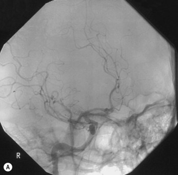
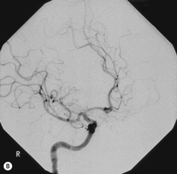
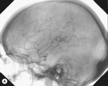
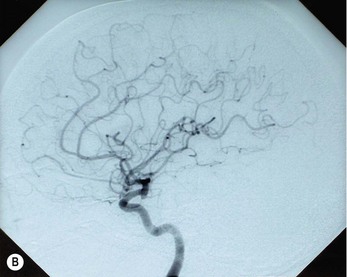
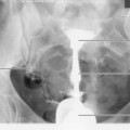
Internal carotid artery (Figs 33.1A,B, 33.2A,B, 33.3A,B)
Typical standard projections are shown in the following table.


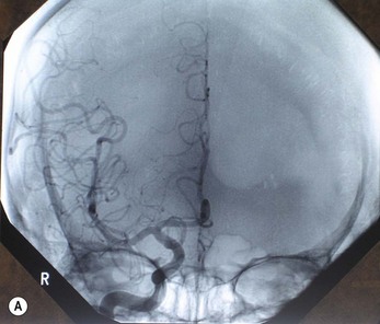
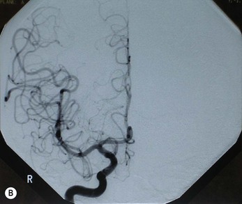
Figure 33.1 OF projection following injection into the right internal carotid artery: (A) unsubtracted; (B) subtracted.


Figure 33.2 AO projection following injection into the left internal carotid artery: (A) unsubtracted; (B) subtracted image.


Figure 33.3 Lateral projection following injection into the left internal carotid artery: (A) unsubtracted; (B) subtracted.

Stay updated, free articles. Join our Telegram channel

Full access? Get Clinical Tree

