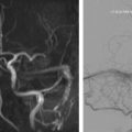
Venous anomalies are the most commonly identified abnormality by imaging in the work-up for pulse synchronous tinnitus. Potential diagnoses include idiopathic intracranial hypertension, sigmoid sinus wall anomalies, transverse and sigmoid sinus stenosis, jugular bulb anomalies, and prominent posterior fossa emissary veins. These causes are discussed in detail along with the association between sigmoid sinus wall anomalies and idiopathic intracranial hypertension.
References
- 1. Vattoth S., Shah R., and Cure J.K.: A compartment-based approach for the imaging evaluation of tinnitus. AJNR Am J Neuroradiol 2010; 31: pp. 211-218
- 2. Mattox D.E., and Hudgins P.: Algorithm for evaluation of pulsatile tinnitus. Acta Otolaryngol 2008; 128: pp. 427-431
- 3. Harvey R.S., Hertzano R., Kelman S.E., et al: Pulse-synchronous tinnitus and sigmoid sinus wall anomalies: descriptive epidemiology and the idiopathic intracranial hypertension patient population. Otol Neurotol 2014; 35: pp. 7-15
- 4. Batra R., and Sinclair A.: Idiopathic intracranial hypertension; research progress and emerging themes. J Neurol 2014; 261: pp. 451-460
- 5. Wall M., Kupersmith M.J., Kieburtz K.D., et al: The idiopathic intracranial hypertension treatment trial: clinical profile at baseline. JAMA Neurol 2014; 71: pp. 693-701
- 6. Degnan A.J., and Levy L.M.: Pseudotumor cerebri: brief review of clinical syndrome and imaging findings. AJNR Am J Neuroradiol 2011; 32: pp. 1986-1993
- 7. Scoffings D.J., Pickard J.D., and Higgins J.N.: Resolution of transverse sinus stenoses immediately after CSF withdrawal in idiopathic intracranial hypertension. J Neurol Neurosurg Psychiatry 2007; 78: pp. 911-912
- 8. Higgins J.N., Cousins C., Owler B.K., et al: Idiopathic intracranial hypertension: 12 cases treated by venous sinus stenting. J Neurol Neurosurg Psychiatry 2003; 74: pp. 1662-1666
- 9. Higgins J.N., and Pickard J.D.: Lateral sinus stenoses in idiopathic intracranial hypertension resolving after CSF diversion. Neurology 2004; 62: pp. 1907-1908
- 10. De Simone R., Marano E., Fiorillo C., et al: Sudden re-opening of collapsed transverse sinuses and longstanding clinical remission after a single lumbar puncture in a case of idiopathic intracranial hypertension. Pathogenetic implications. Neurol Sci 2005; 25: pp. 342-344
- 11. Rohr A., Dörner L., Stingele R., et al: Reversibility of venous sinus obstruction in idiopathic intracranial hypertension. AJNR Am J Neuroradiol 2007; 28: pp. 656-659
- 12. McGonigal A., Bone I., and Teasdale E.: Resolution of transverse sinus stenosis in idiopathic intracranial hypertension after L-P shunt. Neurology 2004; 62: pp. 514-515
- 13. Bono F., Giliberto C., Mastrandrea C., et al: Transverse sinus stenoses persist after normalization of the CSF pressure in IIH. Neurology 2005; 65: pp. 1090-1093
- 14. Connor S.E., Siddiqui M.A., Stewart V.R., et al: The relationship of transverse sinus stenosis to bony groove dimensions provides an insight into the aetiology of idiopathic intracranial hypertension. Neuroradiology 2008; 50: pp. 999-1004
- 15. Schlosser R.J., Woodworth B.A., Wilensky E.M., et al: Spontaneous cerebrospinal fluid leaks: a variant of benign intracranial hypertension. Ann Otol Rhinol Laryngol 2006; 115: pp. 495-500
- 16. Alonso R.C., de la Peña M.J., Caicoya A.G., et al: Spontaneous skull base meningoencephaloceles and cerebrospinal fluid fistulas. Radiographics 2013; 33: pp. 553-570
- 17. Ahmed R.M., Wilkinson M., Parker G.D., et al: Transverse sinus stenting for idiopathic intracranial hypertension: a review of 52 patients and of model predictions. AJNR Am J Neuroradiol 2011; 32: pp. 1408-1414
- 18. Albuquerque F.C., Dashti S.R., Hu Y.C., et al: Intracranial venous sinus stenting for benign intracranial hypertension: clinical indications, technique, and preliminary results. World Neurosurg 2011; 75: pp. 648-652
- 19. Vaghela V., Hingwala D.R., Kapilamoorthy T.R., et al: Spontaneous intracranial hypo and hypertensions: an imaging review. Neurol India 2011; 59: pp. 506-512
- 20. Schoeff S., Nicholas B., Mukherjee S., et al: Imaging prevalence of sigmoid sinus dehiscence among patients with and without pulsatile tinnitus. Otolaryngol Head Neck Surg 2014; 150: pp. 841-846
- 21. Krishnan A., Mattox D.E., Fountain A.J., et al: CT arteriography and venography in pulsatile tinnitus: preliminary results. AJNR Am J Neuroradiol 2006; 27: pp. 1635-1638
- 22. Eisenman D.J.: Sinus wall reconstruction for sigmoid sinus diverticulum and dehiscence: a standardized surgical procedure for a range of radiographic findings. Otol Neurotol 2011; 32: pp. 1116-1119
- 23. Mehanna R., Shaltoni H., Morsi H., et al: Endovascular treatment of sigmoid sinus aneurysm presenting as devastating pulsatile tinnitus. A case report and review of literature. Interv Neuroradiol 2010; 16: pp. 451-454
- 24. Houdart E., Chapot R., and Merland J.J.: Aneurysm of a dural sigmoid sinus: a novel vascular cause of pulsatile tinnitus. Ann Neurol 2000; 48: pp. 669-671
- 25. Sanchez T.G., Murao M., de Medeiros I.R., et al: A new therapeutic procedure for treatment of objective venous pulsatile tinnitus. Int Tinnitus J 2002; 8: pp. 54-57
- 26. Otto K.J., Hudgins P.A., Abdelkafy W., et al: Sigmoid sinus diverticulum: a new surgical approach to the correction of pulsatile tinnitus. Otol Neurotol 2007; 28: pp. 48-53
- 27. Raghavan P., Serulle Y., Gandhi D., et al: Postoperative imaging findings following sigmoid sinus wall reconstruction for pulse synchronous tinnitus. AJNR Am J Neuroradiol 2015; undefined:
- 28. Ong C.K., and Fook-Hin Chong V.: Imaging of jugular foramen. Neuroimaging Clin North Am 2009; 19: pp. 469-482
- 29. Lo W.W., and Solti-Bohman L.G.: High-resolution CT of the jugular foramen: anatomy and vascular variants and anomalies. Radiology 1984; 150: pp. 743-747
- 30. Overton S.B., and Ritter F.N.: A high placed jugular bulb in the middle ear: a clinical and temporal bone study. Laryngoscope 1973; 83: pp. 1986-1991
- 31. Presutti L., and Laudadio P.: Jugular bulb diverticula. ORL J Otorhinolaryngol Relat Spec 1991; 53: pp. 57-60
- 32. San Millan Ruiz D., Gailloud P., Rüfenacht D.A., et al: The craniocervical venous system in relation to cerebral venous drainage. AJNR Am J Neuroradiol 2002; 23: pp. 1500-1508
- 33. Mortazavi M.M., Tubbs R.S., Riech S., et al: Anatomy and pathology of the cranial emissary veins: a review with surgical implications. Neurosurgery 2012; 70: pp. 1312-1318
- 34. Hoffmann O., Klingebiel R., Braun J.S., et al: Diagnostic pitfall: atypical cerebral venous drainage via the vertebral venous system. AJNR Am J Neuroradiol 2002; 23: pp. 408-411
- 35. Lee S.H., Kim S.S., Sung K.Y., et al: Pulsatile tinnitus caused by a dilated mastoid emissary vein. J Korean Med Sci 2013; 28: pp. 628-630
- 36. Forte V., Turner A., and Liu P.: Objective tinnitus associated with abnormal mastoid emissary vein. J Otolaryngol 1989; 18: pp. 232-235
- 37. Lambert P.R., and Cantrell R.W.: Objective tinnitus in association with an abnormal posterior condylar emissary vein. Am J Otol 1986; 7: pp. 204-207
- 38. Chauhan N.S., Sharma Y.P., Bhagra T., et al: Persistence of multiple emissary veins of posterior fossa with unusual origin of left petrosquamosal sinus from mastoid emissary. Surg Radiol Anat 2011; 33: pp. 827-831
Stay updated, free articles. Join our Telegram channel

Full access? Get Clinical Tree


