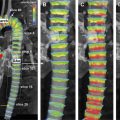
Magnetic resonance assessment of regional myocardial function is a novel potentially important tool for early identification of cardiac pathology. Many cardiac magnetic resonance techniques have been developed for detection and quantification of regional strain abnormalities including steady-state free-precession CINE, tagging, displacement encoding with stimulated echoes, strain encoding imaging, and feature tracking. Potential clinical applications of magnetic resonance strain imaging include early detection of systolic dysfunction in heart failure patients with both ischemic and nonischemic etiologies. Regional function of the myocardium can be quantified using myocardial strain analysis. Myocardial strain is defined as the relative lengthening of the tissue, thus: a normalized measure of deformation. The main parameters used to quantify strain on magnetic resonance imaging are circumferential, longitudinal, and radial strain. Many cardiac magnetic resonance techniques have been developed for detection and quantification of regional strain abnormalities including steady-state free-precession cine, tagging, and displacement encoding with stimulated echoes. Recent clinical studies have shown potential use of these techniques for risk stratification and treatment guidance in patients with congenital and acquired heart diseases. Many cardiac diseases do not affect the heart globally in an early stage. Normal global measures such as ejection fraction can therefore be insensitive to these early regional dysfunctions. Thus, the assessment of the regional myocardium function with magnetic resonance imaging (MRI) poses as a novel potentially important tool for early identification of cardiac pathology. Regional function of the myocardium can be quantified using myocardial strain analysis. Myocardial strain is defined as the relative lengthening of the tissue, thus: a normalized measure of deformation. Cardiac MRI (cMRI) is considered the reference standard for measurement of myocardial strain. Some of the cardiac MRI techniques used for measuring regional myocardial functions are myocardial steady-state free precession (SSFP) cine, tagging, displacement encoding with stimulated echoes (DENSE), strain encoding imaging (SENC), and feature tracking techniques. From a biomechanical point of view, it would be preferable to present the strain given the fiber structure of the heart; however, the complex geometry of the heart makes this challenging. Therefore, the main parameters used to quantify strain on MRI are circumferential, longitudinal, and radial strain ( Fig. 1 ). Another measure used to describe deformation is strain rate, which describes the rate at which the strain is changing over time. Finally, it is possible to quantify diastolic function based on myocardial strain. The main parameter used for quantification of diastolic function is the strain relaxation index (SRI), which is calculated based on the relationship of the circumferential strain with strain rate curves. SSFP has been used routinely in clinical practice for quantification of regional myocardial function. Qualitatively, this technique allows for visual inspection of myocardial thickening during the cardiac cycle in all cardiac regions. Common cardiac planes, named short axis, horizontal long axis, and vertical long axis, provide comprehensive assessment of myocardial contractility in the anterior, lateral, inferior, and septal regions, including the apex, midventricular level, and base of the heart. In addition to visual assessment of cardiac contractility, postprocessing tools allow for quantification of myocardial thickening during the cardiac cycle, which consists of the increase in myocardial wall thickness from end-diastole to end-systole, and is expressed in millimeters. Measurement of wall thickening in the short-axis plane corresponds to the radial strain measurement of the entire transmural myocardium, and can be obtained in each of the 16 American Heart Association (AHA) cardiac segments. Tagging is a cardiac magnetic resonance technique introduced by Zerhouni and colleagues that noninvasively creates markers in the moving tissue in order to observe and assess myocardial motion. Tagging creates visible orthogonal saturation lines (by perturbing the magnetization) that can be imaged and tracked ( Fig. 2 ). Because magnetization is an intrinsic property of the underlying tissue, the tagged lines follow tissue movement, therefore allowing for measurement of the deformation. The main techniques used for strain measurement with tagged lines are based on spatial modulation of magnetization (SPAMM). This technique has been further developed to reduce the fading of the tag lines fading at end-diastole using complementary SPAMM (CSPAMM). The main advantage of this method is its extensive validation studies, including animal and clinical studies. In addition, a range of normal values has already been determined. Disadvantages of the method are mainly related to the fact that the tagged lines fade over time and may not be clearly detected at the end of the cardiac cycle. In addition, postprocessing tools are available, but the process is very time consuming. In addition to conventional tag analysis techniques, a method of extracting myocardial strain measurement from tagging sequences utilizing the tagging frequency called harmonic phase is widely used, and is currently considered the most reliable and reproducible method for postprocessing of tagged images.
Key points
Introduction

Techniques
Steady-State Free Precession Cine
Tagging

Related posts:
![]()
Stay updated, free articles. Join our Telegram channel

Full access? Get Clinical Tree