Chapter 20 Vertebral Augmentation (Vertebroplasty/Kyphoplasty), Transpedicular Approach
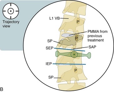
Although alternative techniques have been described, the approach described here involves the use of a trajectory view as the primary approach. The vertebral body is cannulated in the trajectory view (see Appendix 1). Figure 20–10, E, demonstrates the angulation of the needle trajectory in the lateral view with various fracture-pattern scenarios.
A key operational difference between kyphoplasty and vertebroplasty lies in the placement of the working cannula. During a kyphoplasty procedure, when the osteo introducer anchors 1 to 2 mm ventral to the posterior vertebral body (see Figure 20–3, A), a biopsy can be taken; alternatively, the drill can be used to create a path for the balloon tamp (see Figure 20–10, A and B). During vertebroplasty, the introducer cannula is driven from the pedicle toward the midline of the vertebral bone without additional instrumentation. When the tip of the cannula has docked into the posterior vertebral body (see Figure 20–3, A), the visualization of the tip medial to the pedicle is expected in the anteroposterior view; however, advancement should always be done in the lateral view to protect the great vessels.
The oblique transpedicular view is identical to the trajectory view (see Figure 20–1), as described previously.
Note: Please see page ii for a list of anatomical terms/abbreviations used in this book.
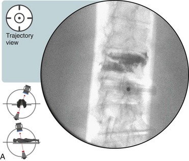
 Transpedicular Advancement: Trajectory View (Figure 20–1)
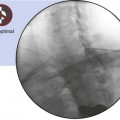
 Trajectory View Safety Considerations


 Confirm the level (with the anteroposterior view).
 Tilt the C-arm to line up the inferior endplate of the vertebral body to be treated.


 Anesthetize the needle tract and periosteum overlying the pedicle.
 Nick the skin (No. 11 blade for kyphoplasty or an 18-gauge needle for vertebroplasty).
 Insert the osteo introducer device or the bone needle, and place the tip onto the pedicle.
 Gently tap the bone needle with a mallet to create a starting hole.
 Advance the needle with gentle manual pressure or mallet tapping.
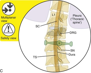
 Multiplanar Views During Transpedicular Advancement (Figures 20-2 and 20-3)
 Anteroposterior View Safety Considerations

Lateral View
Related posts:
 Atlantoaxial Joint Intraarticular Injection
 Lumbar Zygapophysial Joint Nerve (Medial Branch) Radiofrequency Neurotomy, Posterior Approach
 Cervical Interlaminar Epidural Steroid Injection, Paramedian Approach
Stay updated, free articles. Join our Telegram channel

Full access? Get Clinical Tree


