CHAPTER 42 Vestibular Schwannoma
Epidemiology
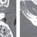
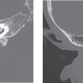
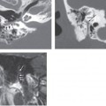
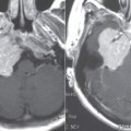
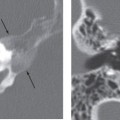
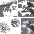
Acoustic neuroma of the eighth cranial nerve (vestibular schwannoma) is, by far, the most common cerebellopontine angle lesion and the most common mass lesion in patients with unilateral sensorineural hearing loss. It arises from the Schwann cells that wrap the vestibulocochlear nerve. It may arise in the internal auditory canal, porus acusticus, or in the internal auditory canal-cerebellopontine angle (IAC-CPA) cistern. It accounts for 8 to 10% of all intracranial tumors and 60 to 90% of all IAC-CPA tumors. Peak age of presentation is 40 to 60 years, though it may be seen in children with neurofibromatosis-2 (NF2). The tumor is slightly more common in females, though intracanalicular tumor may be more common in males. In NF2 patients, acoustic schwannomas are bilateral in 96%.
Clinical Features
Vestibular schwannoma usually presents with unilateral sensorineural hearing loss. Other common presentations include tinnitus, disequilibrium, and decreased speed discrimination. If these tumors are large, then trigeminal or facial neuropathy or even involvement of lower cranial nerves, cerebellar signs, and rarely even hydrocephalus and subarachnoid hemorrhage may occur. The duration of hearing loss before diagnosis is about 3 to 4 years; large tumors with brainstem compression often present with a shorter duration of symptoms and in younger patients.
Pathology
These tumors are slow-growing benign lesions arising from the vestibular portion of the eighth nerve at the oligodendrocyte-Schwann cell junction. Most tumors grow at the rate of 0.02 to 0.2 cm per year. A small percentage of tumors may grow faster, up to 1 cm or more per year.
They are encapsulated and arise eccentrically from the nerve originating in the nerve sheath. Histologically, these lesions are composed of differentiated neoplastic Schwann cells in a collagenous matrix. There are two main histologic spindle cell types: Antoni A tissue, which has a tendency to form palisades; and Antoni B cells, which are less cellular and more loosely arranged and may contain clusters of lipid-laden cells. There is no necrosis, but intramural cysts and rarely hemorrhage within Antoni B tissue may be present. There is strong diffuse expression of S-100 protein.
Treatment
Microsurgery with total resection of small tumors yields good functional preservation of the facial nerve. Hearing preservation is best when the tumor is less than 2 cm and does not invade the internal auditory canal fundus or cochlear aperture. Though there is considerable variation in the approach used, translabyrinthine resection of a large tumor is performed if no hearing preservation is possible. If tumor is intracanalicular, then a middle cranial fossa approach is used, and a suboccipital retrosigmoid approach is used when a cerebellopontine angle component is present. In the case of large acoustic neuromas, subtotal removal and subsequent radiosurgery is one option for maintaining cranial nerve function and long-term tumor growth control.
Stay updated, free articles. Join our Telegram channel

Full access? Get Clinical Tree


