Visual Pathway
At the optic chiasm, the axons from the nasal half of the retina cross to the contralateral optic tract. The axons from the temporal half of the retina, however, do not cross. Thus, in terms of visual fields, the right side (present on the nasal half of the right retina and the temporal half of the left retina) projects to the left hemisphere (optic tract, lateral geniculate ganglion [located in the thalamus], optic radiations, and visual cortex), and vice versa. An optic nerve lesion leads to monocular blindness. An optic chiasm lesion leads to bitemporal hemianopsia (vision loss in the outer halves of both eyes), and an optic tract lesion leads to contralateral homonymous hemianopsia (vision loss on the same side in both eyes).
Any portion of the visual pathway can be involved by an optic glioma, as previously noted. Involvement of the optic chiasm and tract are common. Contrast enhancement of these lesions is variable (from little to prominent). Involvement of the visual pathway is well-identified in these tumors by MR especially on FLAIR scans. As discussed in the brain section, pituitary macroadenomas can extend superiorly, splaying and compressing the optic chiasm, leading to bitemporal hemianopsia.
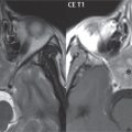
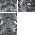
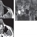
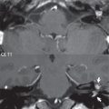
An ophthalmic artery aneurysm is the most common aneurysm to cause visual symptoms, due to impingement on the visual pathway. Ischemia—for example, due to retinal or ophthalmic artery emboli—can lead to transient loss of vision in one eye (monocular blindness). Ischemia involving the visual cortex (occipital lobe) will lead to contralateral homonymous hemianopsia. Optic neuritis can cause either complete or partial loss of vision in one eye, with the most common etiology being multiple sclerosis. Thin-section axial and coronal FLAIR and postcontrast T1-weighted scans (both obtained with fat suppression) are critical for identification of inflammation involving the optic nerve, with edema seen on the former and abnormal contrast enhancement on the latter (typically extending along a relatively long section of the nerve) ( Fig. 2.50 ).

Stay updated, free articles. Join our Telegram channel

Full access? Get Clinical Tree


