Abstract
Widening of the mediastinum is a common observation that may be related to patient body habitus or atherosclerotic dilatation of the aorta and great vessels, but there may also be urgent causes such as aortic dissection or traumatic aortic injury. Less urgent but serious conditions that widen the mediastinum include acute mediastinitis, fibrosing mediastinitis, and tumors, including lymphnoma and metastases.
Keywords
achalasia, aortic dissection, fibrosing mediastinitis, lipomatosis, lymphoma, mediastinal hematoma, mediastinitis, metastases, paraspinal hematoma, traumatic aortic injury, vertebral fractures
Questions
- 1.
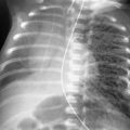
What is the most likely cause of the mediastinal widening in this patient ( Fig 8.1, A and B )?
- a.
Lipomatosis.
- b.
Hematoma.
- c.
Adenopathy.
- d.
Mediastinitis.
- e.
Thymoma.
Fig. 8.1
- a.
- 2.
Which one of the following statements regarding aortic trauma is true?
- a.
The mediastinal width is at least 8 cm.
- b.
Mediastinal hematoma is specific for aortic injury.
- c.
Ascending aorta is the most common sight of aortic injury.
- d.
Computed tomography (CT) angiography is the definitive test for aortic injury.
- e.
Right flail chest excludes aortic injury.
- a.
- 3.
Which of the following statements about mediastinal hematomas are true?
- a.
Often obscure the aortic arch and descending aorta.
- b.
May indicate aortic injury.
- c.
May be caused by vertebral fractures.
- d.
May occur when sternal fractures injure internal mammary vessels.
- e.
All of the above.
- a.
Discussion
Mediastinal widening ( Chart 8.1 ) is a common observation on the posterior-anterior (PA) chest radiograph. It is more difficult to identify confidently on a supine AP view because of magnification and crowding of normal vascular structures by the splinting effect on the patient’s chest. In addition, a lordotic projection distorts and magnifies the superior mediastinum. This is a common problem in the patient who is unable to make a deep inspiratory effort voluntarily, and it is a serious consideration in the emergency department or intensive care unit in which the critically ill patient must be evaluated with portable supine radiography.
- I.
Radiographic technique
- A.
Magnification (anteroposterior [AP] supine chest radiograph, low-volume inspiration)
- B.
Lordotic position
- A.
- II.
Adenopathy
- A.
Neoplasms
- 1.
Lymphoma
- 2.
Primary lung cancer (small cell tumors)
- 3.
Metastases
- 1.
- B.
Inflammatory
- 1.
Mycobacterium avium-intracellulare (in patients with acquired immunodeficiency syndrome [AIDS] 314 )
- 2.
Tuberculosis 261
- 3.
Coccidioidomycosis
- 4.
Anthrax 125
- 5.
Sarcoidosis
- 1.
- A.
- III.
Hematoma
- A.
Aortic injury 655
- B.
Venous and arterial tears
- C.
Sternal fractures
- D.
Vertebral fractures (thoracic and lower cervical spine) 115
- E.
Postoperative bleeding
- F.
Malposition of vascular catheters (also the cause of hydromediastinum)
- A.
- IV.
Vascular structures (nontraumatic)
- A.
Tortuous atherosclerotic dilation of aorta
- B.
Aneurysm
- C.
- D.
Coarctation of aorta 658
- E.
Congenital, left superior vena cava (SVC) with absent right SVC 622
- A.
- V.
Mediastinitis
- A.
Perforated esophagus (Boerhaave syndrome, carcinomas)
- B.
Tracheobronchial rupture (traumatic) 165
- C.
Iatrogenic (postoperative, endoscopic)
- D.
Pneumonias
- E.
Tuberculosis 641
- F.
Coccidioidomycosis
- G.
Histoplasmosis 141 , 492 , 633
- H.
Actinomycosis 393
- I.
Fibrosing or sclerosing mediastinitis 147 , 306 , 371 , 492
- J.
Extension of extrathoracic infections
- 1.
Pharyngeal abscess
- 2.
Abdominal abscess
- 3.
Pancreatitis or pancreatic pseudocyst
- 1.
- A.
- VI.
- A.
Cushing syndrome
- B.
Corticosteroid therapy
- C.
Obesity
- A.
- VII.
Other
- A.
Chylomediastinum (thoracic duct obstruction or iatrogenic laceration) 165
- B.
Mediastinal edema (allergic) 165
- C.
Penetrating trauma (stab wound)
- D.
Achalasia
- A.
Chest radiograph analysis must begin with the identification of as many normal structures as possible, including the ascending aorta, aortic arch, descending aorta, aortic pulmonary window, trachea, paratracheal stripes, carina, main stem bronchi, and paraspinous stripes. 84 Failure to visualize these landmarks requires an explanation and may be an indication for additional procedures, including CT angiography and magnetic resonance imaging (MRI).
Clinical considerations often determine the urgency for a definitive diagnosis. Mediastinal widening after major trauma is strongly suggestive of mediastinal hematoma and requires urgent exclusion of aortic injury. Determining the cause of mediastinal widening in patients without a history of trauma may also be challenging.
Determining the cause of the mediastinal widening is often more difficult than recognizing the presence of an abnormality, especially in older patients with atherosclerotic vascular disease that leads to dilation and tortuosity of the aorta and great vessels. Patients who have had cardiac or vascular surgery are easily recognized by the radiographic identification of surgical clips and metal sutures, but this may further confuse the evaluation of mediastinal widening. These patients may have both tortuous vessels and postoperative abnormalities, including hematomas during the acute convalescent period and, later, mediastinal scarring. A mass lesion that develops subsequent to mediastinal or cardiac surgery may be obscured by postoperative changes.
Adenopathy
Mediastinal tumors are expected to produce discrete masses, but neoplasms that involve multiple lymph node groups may cause diffuse mediastinal widening. This type of tumor dissemination results from lymphomas and metastases from either primary lung cancer or distant primaries. It is particularly common with poorly differentiated primary tumors, such as small cell lung cancer. Such extensive nodal involvement may also be seen with metastases from retroperitoneal tumors and even from testicular tumors, particularly seminomas.
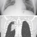
Lymphoma frequently involves mediastinal lymph nodes and, when the involvement is extensive, numerous large nodes may diffusely widen the mediastinum ( Fig 8.2, A and B ). The tumor should regress following chemotherapy, but the nodes may be replaced by extensive fibrosis, which may leave residual mediastinal widening. This is most often recognized by the identification of paramediastinal reticular opacities that result from mediastinal fibrosis. In their early stages, radiation pneumonitis and fibrosis may appear to become more prominent over a short period, requiring further consideration of recurrent tumor. Increasing opacity from the radiation effect on the mediastinum may be further evaluated with MRI, which has been reported to be useful for identifying viable tumor in the midst of postradiation scarring.