![]() To demonstrate the optimum position for patient, examiner, and equipment to avoid work-related injury.
To demonstrate the optimum position for patient, examiner, and equipment to avoid work-related injury.
![]() To introduce the most common, universally applied transducer manipulation techniques employed during scanning.
To introduce the most common, universally applied transducer manipulation techniques employed during scanning.
KEY TERMS
Anchoring the transducer
Heel-toe manipulation
Negative pressure
Neutral position
Positive pressure
Probe angulation
Probe rotation
Probe translation
INTRODUCTION
Ultrasound scanning is a combination of skill, science, and art. There is a definite set of sonographic techniques that require knowledge and discipline which, when mastered, apply to all areas of scanning. Sonography is unlike other forms of medical imaging in several ways. Primarily, the scanning process is diagnostic in nature, requiring each examination to be customized to the patient in response to observations and clinical assessment. Unlike radiography or computed tomography, in which rigid protocols are prescribed based on patient history and are not varied during the examination, sonography is adaptable in its execution in response to findings. The sonographer is first an investigator. Information is acquired by exploration, comparison with known normal anatomy and variations thereon, and recognition of abnormal results. Sonography requires a thorough understanding of instrumentation, physics, anatomy, physiology, and the limitations imposed by the combination of these. The knowledge of how to work around these limitations is essential in order to obtain the desired diagnostic information. The examiner must be able to spend as much time as necessary to conduct a comprehensive examination, without strain or fatigue.
Foremost in importance for the sonographer is the development of good habits that minimize strain on the back, arms, wrists, neck, and shoulder joints. A publication issued by the NIOSH in 2006 examined the causes of work-related musculoskeletal injuries in sonographers and included recommendations to help prevent such injuries.1 The report stated, in part, that injuries resulted from (1) static and awkward postures and movement resulting from the use of the transducer and positioning of both patients and equipment; (2) persistent and continual pressure for sustained periods of time during exams; (3) poor workplace ergonomics in the design of equipment, chairs, tables, and lighting; and (4) increased exam scheduling. Consideration of these factors is even more imperative for users of portable scanners. Most of the larger ultrasound systems have incorporated ergonomic features such as mobility of the monitor and keyboard, devices to assist in cable control, and other equipment designs that are not present on the portable scanners.
Another study by Evans, Roll, and Baker in 2009 surveyed 5200 sonographers and vascular technologists.2 Of the 2963 who responded, 90% reported that they are currently working in pain. Shoulder pain is reported as being most common, with older and more experienced sonographers having more finger, hand, and wrist pain than other groups. Pain continues to be related to pressure applied to the transducer, abduction of the arm, and twisting of the neck and trunk. Ergonomics is an essential component in the practice of ultrasound, both in terms of the long-term health of the sonographer and the ability to conduct the optimal examination.
SCANNING POSITION
Whether a seated or a standing position is utilized by the sonographer, he/she must establish a position where movement and placement of the transducer is controlled absolutely in order to acquire the highest quality diagnostic images. In addition, this scanning posture should be achieved without strain to the sonographer’s back, legs, neck, arms, or hands. The goal is for the sonographer’s body to be in a neutral position while scanning.
Seated Position
The seated position is always preferable if circumstances permit. A scanning room may be configured to allow sonographers to sit while scanning the patient. Ideally, the height of the scanning table and the chair height are adjustable. Dedicated scanning tables and chairs are not inexpensive, but well worth the extra cost in the form of fewer sonographer injuries and less loss of staff work hours. Chair height and table height are critical in order to enable the sonographer to rest his/her scanning arm upon the patient or some other support (Figure 9-1).
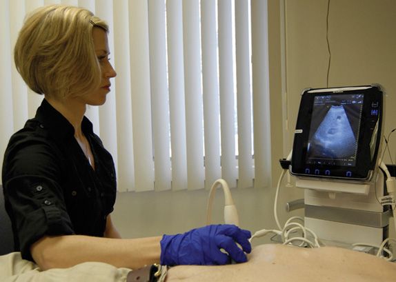
FIGURE 9-1. Seated sonographer with proper chair height and arm support.
Standing Position
If a standing position is necessary, the table height is the major consideration. Again, the proper table height is critical to allow the sonographer to rest the scanning arm. If the sonographer cannot manage a neutral position and must lean over while scanning, back and shoulder problems will arise.
SCANNING TABLES
In the “old days,” obtaining an ultrasound scanning table was simple—just locate a patient litter or stretcher that was not in use. As tempting as that choice may be for the cost-conscious, these devices have been, over the years, a sure formula for sonographer injury. The scanning table should be adjustable in height, be movable, and have lockable wheels. The large bumpers and railings on the sides of patient stretchers prevent a patient from being close to the sonographer. To compensate for the increased separation, the sonographer must bend over to reach the patient. Another poor choice of scanning table is the standard patient exam table found in most physicians’ offices. The patient exam table has a fixed height and is difficult for the sonographer to approach closely enough to avoid having to lean and reach. Dedicated scanning tables for various applications as well as adjustable chairs and other ergonomically correct devices are available from a number of manufacturers.
PATIENT POSITION
The sonographer should begin each exam by positioning the patient correctly on the table. The extra few seconds this step requires will be a significant benefit to sonographer in terms of comfort and safety. Positioning or repositioning of the patient must be incorporated into the individual sonographer’s workflow to be effective. Moving the patient closer to the examiner by only a few inches can eliminate the need for reaching and bending while scanning (Figures 9-2 and 9-3). Asking a patient to slide an inch or two toward the head or feet can also positively affect the sonographer’s body position (Figures 9-4 and 9-5).
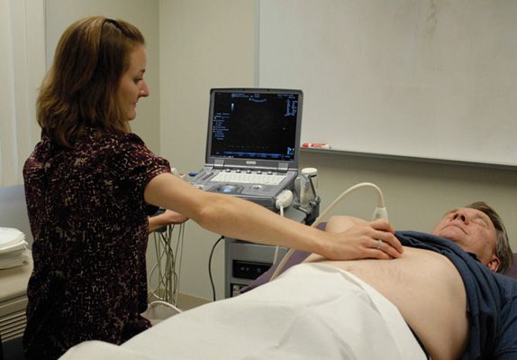
FIGURE 9-2. Seated sonographer in an awkward position having to lean to reach the patient. Observe the absence of arm support.
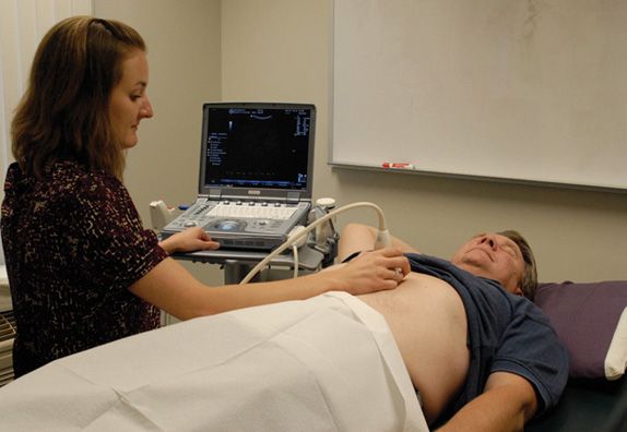
FIGURE 9-3. Patient moved closer to the table edge allowing the sonographer’s body and scanning arm to remain in a neutral position with proper arm support.
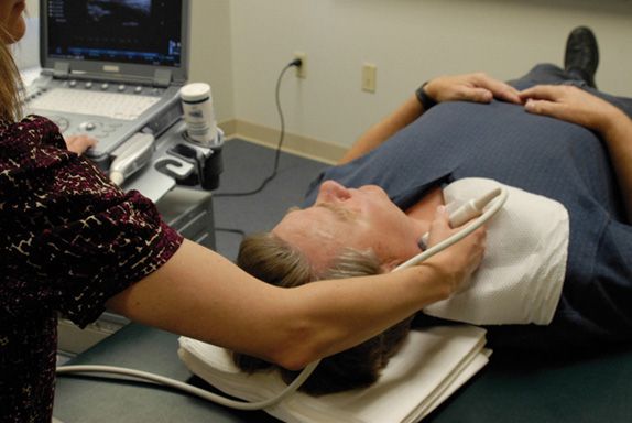
FIGURE 9-4. A carotid scan with patient positioned too far toward the foot of the table causing the sonographer to reach uncomfortably.
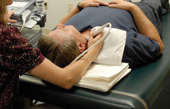
FIGURE 9-5. Patient repositioned a few inches closer to the head of the table with the sonographer’s arm now well supported and her body in a neutral position.
EQUIPMENT SUPPORT PLATFORM
Because portable scanners are easily movable and fold up like a laptop computer, many users assume that they can be placed and operated anywhere there is an available level surface. Portable scanners have been placed on bedside tables, on ECG carts, or next to the patient on the exam table. Sometimes the level surface is formed by holding the device in the lap of the sonographer. All of these are poor choices, both from the point of stability of the instrument and accessibility by the examiner. In addition, care must be taken to provide safe and positive support for the transducer and cable, both of which are susceptible to damage from dropping or pinching. Transducer cables are typically the most vulnerable components of the system. A damaged cable often makes the entire transducer assembly unusable and irreparable. If two or more transducers are available for use with the system, equal care must be taken to protect the transducer(s) that are not currently active, including their cables and connectors. An inadequate platform for the equipment also makes it more difficult for the sonographer to access the system controls and view the monitor without discomfort.
The portable scanner should be adequately supported in a position in which the sonographer is able to operate the controls with his/her free hand (non-scanning hand) without reaching, bending, or twisting. The screen should be positioned such that the sonographer’s head is not turned at an acute angle for viewing.
Most manufacturers of mid- to high-end portable units also offer a mobile equipment cart as an option with the system. These carts are typically designed for the specific model of scanner, and most are light enough in weight that they can be picked up and placed into a vehicle trunk or cargo area if the scanner is to be transported to different locations. Although these carts are certainly more expensive than a bedside table, they provide portability, ease of use, and protection of valuable transducers and cables. Figure 9-6 shows examples of portable system carts available from three manufacturers.
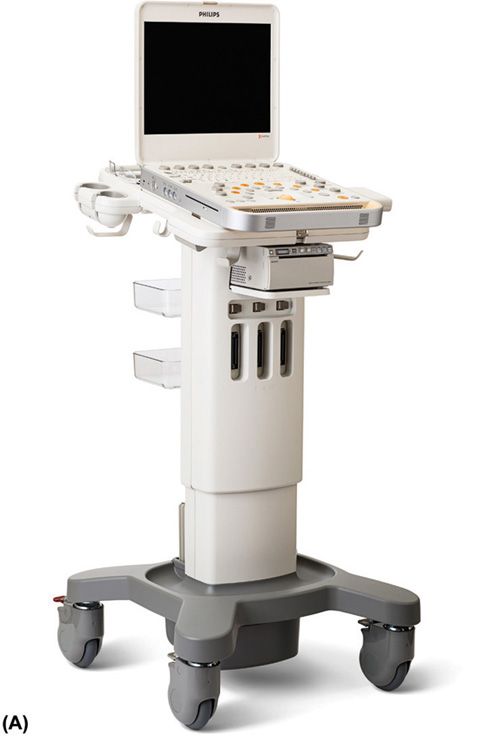

FIGURE 9-6. Portable equipment carts. (A) Philips CX-50 (Courtesy of Philips Healthcare). (B) Sonosite Edge (Courtesy of FujiFILM-Sonosite, Inc.) (C) GE Loqiq-e (Courtesy of GE Healthcare).
EQUIPMENT POSITION
The portable scanner, when adequately supported on a mobile cart, can be repositioned as needed during the course of an ultrasound examination. In order to maintain a neutral posture throughout the examination, especially one like a lower extremity venous evaluation, the sonographer should be able to roll the chair and machine along the table as scanning progresses from the patient’s torso region toward the feet. Here again, the mobile equipment cart provided by the manufacturer provides the most flexibility, while assuring adequate protection of transducers and dangling cables (Figure 9-7).
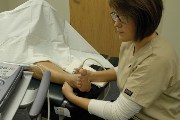
FIGURE 9-7. Seated sonographer performing venous lower extremity exam with machine pulled toward the patient’s feet.
SCANNING ARM SUPPORT
Related posts:

Stay updated, free articles. Join our Telegram channel

Full access? Get Clinical Tree


