Tissue Suppression Techniques
Introduction
One of the beauties of MR, especially with some of the new features, is the ability to image a body part while “suppressing” the signal coming from a certain selected tissue. This suppression allows perturbation of tissue contrast to enhance the signal coming from tissues of greater interest (such as a pathology). Two types of tissues are commonly suppressed in clinical practice: fat and water.
Suppression Techniques
Several suppression techniques are available, some of which are listed below.
Inversion recovery techniques
Chemical (or spectral) saturation or frequency-selective presaturation
Spatial presaturation in the field of view (FOV)
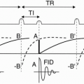
Inversion Recovery (IR) Techniques. This technique was discussed at length in Chapter 7. The IR pulse sequence diagram (PSD) is shown in Figure 25-1. By appropriate selection of TI (time to inversion), we can nullify or suppress a certain tissue. In fact, as we saw in Chapter 7, if TI = (ln 2) [T1(tissue x)] = 0.693 T1(tissue x) then tissue x is nulled. Thus, TI can be chosen to null fat or water or any other desired tissue depending on the application (Fig. 25-2).
Example
What is the TI used in STIR? At 1.5 T, T1 of fat is approximately 200 msec. Then TI = 0.693 × 200 ≅ 140 msec FLAIR (Fluid-Attenuated Inversion Recovery). This is an IR technique that nulls fluid. For example, this sequence is used in the brain to suppress cerebrospinal fluid (CSF) to bring out the periventricular hyperintense lesions, such as multiple sclerosis (MS) plaques (Fig. 25-3).
Example
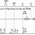
What is the TI used in FLAIR? At 1.5 T, T1 of CSF is approximately 3600 msec. Then TI = 0.693 × 3600 ≅ 2500 msec Fast FLAIR. STIR sequences are typically performed with a fast spin-echo (FSE) technique; however, there are some novel changes that must be performed to apply FSE technique to the normally slow FLAIR sequence and achieve CSF suppression in a fast manner.1 Figure 25-4 depicts a schematic representation for Fast FLAIR. In this scheme, the following parameters are used:
 Figure 25-1. Inversion recovery (STIR shown). The recovery curves following the 90° and 180° pulses are illustrated for fat and water. |
In Figure 25-4, there are two packets of 15 slices each. During the first 5000 msec, 15 slice-selective 180° inverting pulses are followed 2500 msec (TI) later by 15 slice-selective FSE readout in 2500 msec. During the second 5000 msec, recovery occurs in the first 15 slices (for a total TR of 10,000 msec), and the process is repeated on the second set of 15 interleaved slices. In all, 30 slices are acquired.
A TR of 10,000 msec allows almost complete longitudinal recovery of CSF. This relatively long time period also makes it possible to perform multislice interleaving during inversion and readout. The inversion “period” is the time during which 15 slice-selective 180° pulses are applied. It is the time TI (2500 msec in this case) from the first 180° inverting pulse to the 90° pulse at the beginning of the readout period. FSE readout takes 136 msec (8 × 17 msec) for each slice. The readout “period” is the time (also 2500 msec) during which 15 sliceselective readouts are performed. The recovery period is the time from the slice-selective 90° pulse
beginning an FSE readout to the next slice-selective 180° inverting pulse, which is TR–TI (10,000 − 2500 = 7500 msec in this case).
beginning an FSE readout to the next slice-selective 180° inverting pulse, which is TR–TI (10,000 − 2500 = 7500 msec in this case).
 Figure 25-2. TI required to null fat, brain, or CSF. |
 |
 Figure 25-4. A schematic representation for Fast FLAIR. There are two packets of 15 slices each. During the first 5000 msec, 15 slice-selective 180° inverting pulses are followed 2500 msec (TI) later by 15 sliceselective FSE readout, in 2500 msec
Get Clinical Tree app for offline access
Related posts:Stay updated, free articles. Join our Telegram channel
Full access? Get Clinical Tree


|