![]() To understand the different capabilities of handheld versus portable console scanners.
To understand the different capabilities of handheld versus portable console scanners.
![]() To recognize the equipment requirements for various sonographic applications.
To recognize the equipment requirements for various sonographic applications.
KEY TERMS
Hand-held scanner
Portable console scanner
Scanning window
Transducer Footprint
INTRODUCTION
Requirements for portable ultrasound equipment are based primarily on the proposed applications. The cost of point-of-care scanners ranges from around $6000 for a portable “hand-held” scanner to well over $100,000 for a full-featured laptop system. The purchaser must match the capabilities (and price) of the point-of-care system with the expected clinical use. If the objective is to provide a high-end, full-range diagnostic service capable of cardiac, obstetrics, abdominal, breast, and musculoskeletal (MSK) imaging, a low-cost scanner will not be adequate.
TRANSDUCER APPLICATIONS
In Chapter 3, six types of transducers have been discussed. Each transducer type is intended for specific applications and has unique features and image formats. The scanner must accommodate not only the transducer itself, but also the electronics to support that transducer. For example, a phased sector transducer requires different hardware and electronics than a linear array transducer. Essentially, the design of an ultrasound unit is based on the intended transducers of use. Transducers can be grouped more generally into two main categories: (1) sector type and (2) linear array.
Sector-Type Transducers
The sector (or phased array) transducer generates a cone-shaped image format with the sound source originating from a small apex. The area of the transducer face that contacts the patient is called the transducer footprint. A small transducer footprint is advantageous for applications for which there is very limited access at the skin surface. The most common use of sector transducers is imaging of the heart. Sound enters the body via a small scanning window and the two-dimensional field of view widens with scan depth. Indeed, most cardiac scanning would not be possible without sector technology. The phased array is also utilized for intercostal scanning within the abdomen.
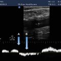
Sector transducers are required for adult and pediatric cardiac scanning, but also can perform abdominal, gynecological, and basic obstetrical scanning reasonably well. Sectors are not suitable for musculoskeletal, breast, or thyroid (small parts) imaging because field of view is narrowed in the near field and maximum frequency is generally limited to 5 MHz. They are also inadequate for most vascular applications. Cardiac applications ideally require two transducers with different frequency ranges for both adult and pediatric use. The majority of hand-held units utilize sector technology (Figure 7-1).
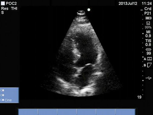
FIGURE 7-1. Sector image of heart, four-chamber view, obtained with a hand-held scanner.
Linear Array Transducers
Linear array transducer technology encompasses multiple transducer types including the straight (flat) linear array, convex or curvilinear array, steered linear array, and vector array. These transducer types rely primarily on the same basic method for image formation, that is, the elements are excited in successive groups across the face of the transducer to create the two-dimensional image. The curvilinear array, with its curved geometry, produces a wider field of view than does a standard linear array. The vector array extends the width of the field of view by implementing phased beam steering (similar to a phased array) at the periphery. The steered linear array is capable of steering (angling) the entire image area to the left or right when necessary.
The linear array is the transducer of choice for obstetrics, gynecology, breast, superficial structures including MSK, and vascular applications (if duplex spectral Doppler is available). Abdominal scanning with a curvilinear array is now the standard of practice and this transducer can also achieve intercostal scanning of the abdomen (Figures 7-2 and 7-3). At least three separate broadband linear array transducers are necessary to perform the full complement of clinical applications: a low-frequency curvilinear array in the range of 3–5 MHz (abdomen and OB-gyn), a 7- to 10- MHz transducer with Doppler capability (vascular), and a high-frequency linear array in the 12–15 MHz range (breast, thyroid and MSK) (Figure 7-4). In addition, a dedicated OB-gyn scanner requires an endovaginal transducer as well as three-dimensional (3D) technology for obstetrical imaging. Linear array transducers are not suitable for cardiac imaging due to their large transducer footprint.
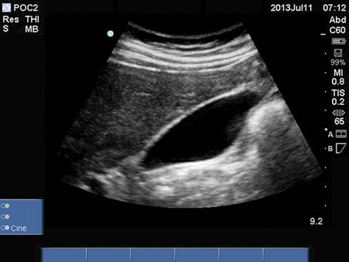
FIGURE 7-2. B-mode image of the gallbladder acquired with a curvilinear array.
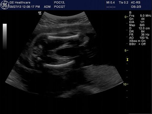
FIGURE 7-3. B-mode image of a fetus showing both femurs acquired with a curvilinear array.
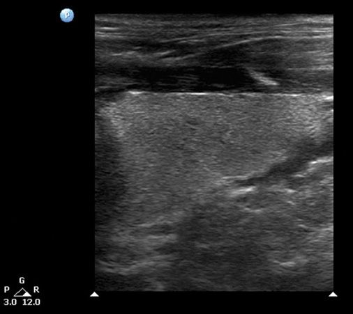
FIGURE 7-4. B-mode image of the thyroid gland acquired with linear array.
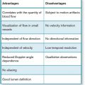
EVALUATION OF BLOOD FLOW
Related posts:


Stay updated, free articles. Join our Telegram channel

Full access? Get Clinical Tree


