19 Tendons
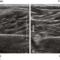
Tendons are the strong structures that connect muscle to bone. Their fibrillar echotexture (fiber-like, appearing as the fine hairs of a violin bow) results from parallel collagen bundles. Because of this ordered architecture, tendons are highly anisotropic, meaning that the received echoes are highly dependent on the angle of insonation.1–3
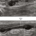
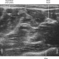
Tendons and nerves are both imaged during regional block procedures, and therefore some commentary regarding their discriminatory features is appropriate. Although the two structures can appear similar, tendons and nerves are primarily distinguished by tracing their course. Because tendons only form at the ends of muscle, changes in cross-sectional area along the course are substantial. For nerves, the cross-sectional area is relatively constant along the nerve path. In addition, the amplitudes of the received echoes from tendons are more sensitive to transducer inclination than nerves (tendons are more anisotropic than nerves). At high frequencies of insonation (≥10 MHz), the fibrillar echotexture of tendons can be distinguished from the fascicular echotexture of nerves (Table 19-1).4
Table 19-1 Characteristics of Nerves and Tendons that Can Be Identified with Ultrasound Imaging
| Characteristic | Nerve | Tendon |
|---|---|---|
| Echotexture | Fascicular | Fibrillar |
| Elemental composition | Fascicles (coarse, thick, wavy, less numerous) | Fibrils (fine, thin, straight, more numerous) |
| Internal architecture | Plexiform (combine and divide) | Parallel fibrils |
| Cross-sectional area | Constant along nerve path | Forms at the ends of muscle |
| Overall shape | Round, oval, or triangular |