37 Ulnar Nerve Block
The ulnar nerve is a branch of the medial cord of the brachial plexus. The ulnar nerve provides sensation of the dorsal and palmar sides of the ulnar aspect of the hand. It leaves the neurovascular bundle in the axilla to travel through the cubital tunnel. In the forearm it joins the ulnar artery on its medial side. The ulnar nerve usually lies between the ulnar artery and the flexor carpi ulnaris (FCU) tendon in the forearm. The dorsal cutaneous branch leaves the ulnar nerve in the forearm proximal to the wrist.1,2 At the level of the hamate, the ulnar nerve divides into its superficial sensory branch and its deep motor branch.
Suggested Technique
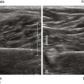
The ulnar nerve is usually blocked just proximal to its juncture with the ulnar artery in the forearm.3 In this location the nerve is either oval or triangular. The block is performed with the patient supine and the arm supinated. The needle tip is placed within the fascial plane that connects the ulnar nerve and ulnar artery using an in-plane approach from the lateral side of the forearm. To access this plane with the block needle it is best to puncture the fascia and slowly inject as the needle is pulled back.
A relatively common (3%-10%) anatomic variant is superficial ulnar artery, whereby the ulnar artery lies superficial to the flexor muscles.4