65 Pneumothorax and Other Chest Pathology
Ultrasound imaging can prevent and diagnose pneumothorax. Chest sonography is therefore an essential skill for those performing regional blocks. Regional anesthesia is the leading cause of pneumothorax during anesthesia, and twice as likely as the next leading cause.1 This is particularly surprising given that line placement is another potential cause of perioperative pneumothorax.
The incidence of pneumothorax after traditional supraclavicular block has been reported to be as high as 0.5% to 6%.2 This high incidence has led to concern that supraclavicular blocks should not be performed on outpatients. Pneumothorax after ultrasound-guided regional block has been reported.3
Several risk factors for pneumothorax and chest tube placement have been identified in patients undergoing interventional procedures.4 These include the needle traversing aerated lung or a lung fissure, lung hyperinflation from chronic obstructive lung disease, and positive-pressure ventilation.
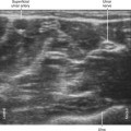
Because of the marked acoustic impedance mismatch with soft tissue, the pleura generates a brighter echo than the surface of the first rib. Comet-tail artifact can be observed deep to strongly reflecting structures, such as the lung.5,6
Related posts:


Stay updated, free articles. Join our Telegram channel

Full access? Get Clinical Tree


