Interscalene and Supraclavicular Blocks

27 Interscalene and Supraclavicular Blocks


The first use of ultrasound to guide regional block was for brachial plexus block above the clavicle.1 These pioneers used an offline Doppler technique to mark the position of the subclavian artery as a surrogate landmark of the brachial plexus. Although today there may be many criticisms of this technique, their results were impressive: a 98% success rate with no complications.


Ultrasound imaging blurs the distinction between interscalene and supraclavicular blocks. If the brachial plexus is seen stacked between the anterior and middle scalene muscles, the block is generally referred to as an interscalene block. If the brachial plexus is seen as a compact group of nerves lying superior and lateral to the subclavian artery, the approach is generally referred to as a supraclavicular block. Because this distinction can be subtle, both blocks will be treated together.


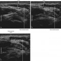
Variations in brachial plexus anatomy with respect to the scalene muscles are common. The cephalad components of the plexus (in particular, the C5 and C6 ventral rami) often pass over or through the anterior scalene muscle. This may pose a problem for nerve stimulation–based approaches to brachial plexus blocks above the clavicle. The incidence of scalene muscle anomalies is similar for sonography of volunteers and in cadaveric dissections, suggesting that ultrasound can accurately detect these anomalies.2


Cervical ribs are relatively uncommon, occurring in about 0.5% of the population.3,4 Most cervical ribs are partial (incomplete) and therefore do not pose a problem. However, if the cervical rib is sufficiently large, transducer manipulation can be difficult, and acoustic shadowing by the bone obscures imaging of the brachial plexus.


Suggested Technique


The monofascicular ventral rami of the brachial plexus are hypoechoic and therefore can be difficult to identify between the scalene muscles. The ventral rami can be similar to blood vessels in their ultrasound appearance. The brachial plexus lies deep to the tapering posterolateral edge of the sternocleidomastoid in the neck.


The best nerve visibility is usually near the first rib because the brachial plexus is compact and lateral to the subclavian artery. The plexus contains more connective tissue moving from interscalene to supraclavicular views, resulting in more hyperechoic echotexture. To obtain a good supraclavicular view with the subclavian artery in true short axis, the imaging plane must face caudally at the brachial plexus (not posteriorly). Brachial plexus imaging in the supraclavicular region is most consistent and can be used to trace the plexus back to the interscalene groove. Perform the block where the imaging is most reliable.


The semisitting (beach-chair) position helps comfort the patient, lowers the arm by gravity, and brings the plane of imaging closer to the plane of the display. The head-of-bed elevation should be about 45 degrees. The patient’s head turns to the opposite side from the block. The operator stands either at the head of the bed or at the side of the bed, depending on the side of the block and the handedness of the operator.


Working room is limited above the clavicle, and therefore a compact transducer is favored. A small curved or small linear (20- to 25-mm footprint) transducer is preferred. The compact transducer can be rocked back to improve needle visibility. Broad linear probes are more difficult to rock than narrow linear or curved transducers for this procedure. The ulnar aspects of both hands of the operator are placed on the patient for the best control of the needle and transducer.


A short (50 mm), broad (21 gauge) echogenic needle is used for optimal control and visibility. Hand-on-needle hub (not hand on syringe) is recommended for better needle control. A medial to lateral in-plane needle path heads away from pleura.


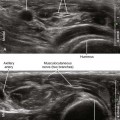
Most authors recommend a multiple injection technique to ensure complete plexus anesthesia. With this approach the initial aim of the needle is deep (under the more caudal elements of the plexus) so that the brachial plexus rises closer to the skin surface with the injection of local anesthetic. This makes the subsequent needle passes easier to perform. The needle tip should be positioned adjacent to the components of the brachial plexus for injection within the interscalene groove.5

Only gold members can continue reading. Log In or Register to continue

Stay updated, free articles. Join our Telegram channel

Mar 5, 2016 | Posted by in ULTRASONOGRAPHY | Comments Off on Interscalene and Supraclavicular Blocks

Full access? Get Clinical Tree

Get Clinical Tree app for offline access