Case 10(Continuation of Case 9)
Indication: Status 2 years after excisional biopsy (benign) for suspected cancer. New lump near scar.
History: Unremarkable.
Risk profile: Breast cancer in grandmother at the age of 82 years.
Age: 54 years.
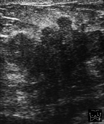
Fig. 10.1 Ultrasound image from the area of the palpable mass.
Clinical Findings
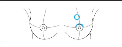
2 cm resistance in the upper inner quadrant of the left breast in the region of the previous open biopsy. Uncomplicated scar.
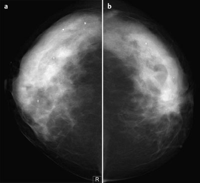
Fig. 10.2a,b Digital mammography, CC view.
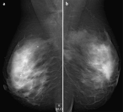
Fig. 10.3a,b Digital mammography, MLO view.
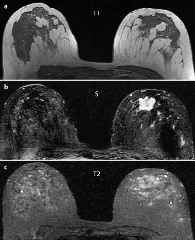
Fig. 10.4a–c Contrast-enhanced MR mammography.
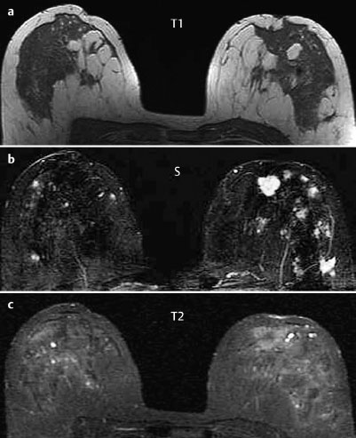
Fig. 10.5a–c Contrast-enhanced MR mammography.
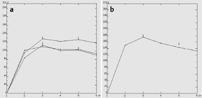
Fig. 10.6a, b Signal-to-time curves of the lesion in the left breast.
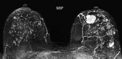
Fig. 10.7 Contrast-enhanced MR mammography. Maximum intensity projection.
| Please characterize ultrasound, mammography, and MRI findings. What is your preliminary diagnosis? What are your next steps? |
Stay updated, free articles. Join our Telegram channel

Full access? Get Clinical Tree


