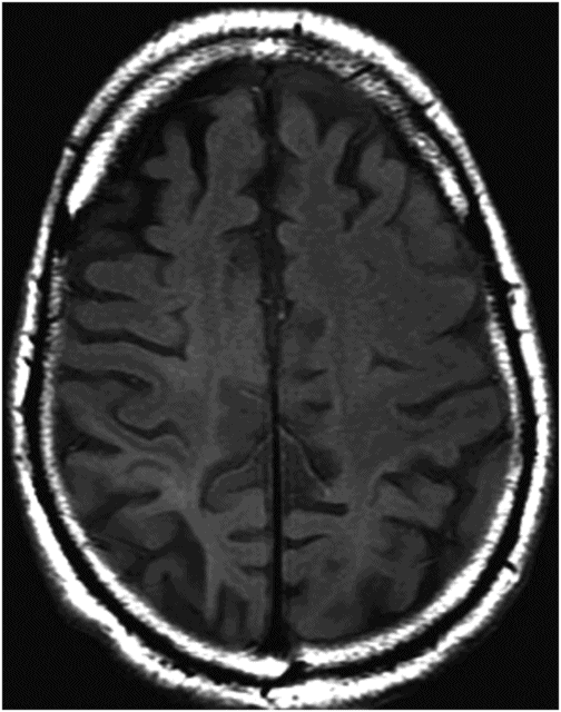
Axial FLAIR image through the centrum semiovale.
Corticobasal Degeneration
Primary Diagnosis
Corticobasal degeneration
Differential Diagnoses
Progressive supranuclear palsy
Alzheimer disease
Posterior cortical atrophy
Stroke
Creutzfeldt-Jakob disease
Frontotemporal lobar degeneration
Imaging Findings
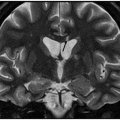
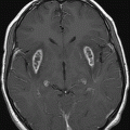
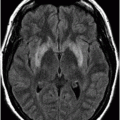
Fig. 10.1: Axial FLAIR image through the centrum semiovale demonstrated bilateral posterior frontal and parietal atrophy as evidenced by secondary enlargement of the sulci and predominantly right-sided thinning of the gyri. Fig. 10.2: Axial FLAIR image through the centrum semiovale (obtained inferior to Fig. 10.1) demonstrated mild, posterior-predominant atrophy, in addition to abnormal FLAIR signal in both posterior parietal lobes, as well as in the right posterior frontal lobe.
Discussion
The clinical description of this case is typical of corticobasal syndrome (CBS). Imaging demonstrates posterior-predominant atrophy involving both the parietal lobes and the right posterior frontal lobe. Combined clinical and radiologic findings are suggestive of CBS, likely associated with Alzheimer disease (AD).
No focal encephalomalacia was detected, ruling out large-artery distribution stroke. The clinical course of Creutzfeldt-Jakob disease features a rapidly progressive dementia with or without myoclonus typically followed by death within the same year. Our patient was symptomatic for more than two years. Posterior cortical atrophy is a radiologic description of posterior-predominant brain atrophy and can be seen in many different neurodegenerative diseases including CBS. Although progressive supranuclear palsy (PSP) and AD are two distinct diseases, both of these conditions can present clinically with CBS. Therefore, it is possible that even though the patient has CBS, he/she will have pathologic diagnosis of corticobasal degeneration (CBD), PSP, or AD (see the discussion below).
Corticobasal syndrome is a rare, progressive neurodegenerative disease with a heterogeneous clinical presentation consisting of motor, sensory, behavioral, and cognitive symptoms that frequently overlap with other neurodegenerative disorders such as frontotemporal lobar degeneration (FTLD), AD, and PSP. The classical clinical manifestation of CBD is known as corticobasal syndrome (CBS) and is characterized by levodopa-unresponsive Parkinsonism, progressive asymmetric rigidity, apraxia, aphasia, and dystonia, which can be found in up to 50% of pathologically proven cases of CBD (CBS-CBD). However, CBS can be a presenting symptom of Alzheimer disease (CBS-AD), progressive supranuclear palsy (CBS-PSP), and frontotemporal lobar degeneration (CBS-FTLD) making its diagnosis difficult.
Typically, CBD demonstrates asymmetric focal atrophy of the frontoparietotemporal lobes, sparing the brainstem and cerebellum; however, the substantia nigra may demonstrate loss of pigmented neurons. Manifestation of brain atrophy in CBS is heterogeneous and condition-dependent such as frontotemporal atrophy in CBS-FTLD, parietotemporal atrophied CBS-AD, and superior posterior frontal in CBS-CBD/CBS-PSP. Predominant dominant frontal lobe atrophy is seen in CBS-FTLD, whereas predominant dominant parietal lobe atrophy is seen in CBS-FTLD and CBS-AD. Predominant cortical atrophy is not a feature of CBS-CBD and CBS-PSP.
Corticobasal degeneration is a 4R-tauopathy similar to PSP (Part I: Case 11). On histopathology, there are neuronal spherical corticobasal bodies that are tau-4R immunoreactive, and are found mostly in the frontal and parietal cortex. However, the histopathologic hallmark of CBD is the presence of numerous Gallyas – and tau-immunoreactive neuronal threads/astrocytic plaques – in the neuropil of affected gray matter and adjacent white matter.
Stay updated, free articles. Join our Telegram channel

Full access? Get Clinical Tree


