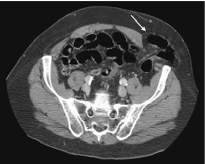
CASE 101 A middle-aged patient complains of a palpable abdominal wall mass. Fig. 101.1 Axial noncontrast CT image through the pelvis demonstrates a defect in the anterior abdominal wall, just lateral to the semilunar line. The sigmoid colon is seen herniating through this defect (arrow). Axial noncontrast computed tomography (CT) image through the pelvis demonstrates a defect in the anterior abdominal wall, just lateral to the semilunar line. The sigmoid colon is seen herniating through this defect (Fig. 101.1). Spigelian hernia
Clinical Presentation
Radiologic Findings
Diagnosis
Related posts:


Stay updated, free articles. Join our Telegram channel

Full access? Get Clinical Tree


