CASE 103 A 73-year-old woman presents with abdominal discomfort and flank pain. Fig. 103.1 Coronal reformatted contrast-enhanced CT image show a heterogeneous, ill-defined mass in the left perinephric region surrounding the left kidney. The lesion shows areas of fat density and foci of calcifications adjacent to the left kidney. Coronal reformatted, contrast-enhanced computed tomography (CT) image shows a heterogeneous, ill-defined mass in the left perinephric region surrounding the left kidney. The lesion shows areas of fat density and foci of calcifications adjacent to the left kidney (Fig. 103.1). Dedifferentiated retroperitoneal liposarcoma Liposarcoma is the second most common type of soft tissue sarcoma in adults and is most commonly seen in the 5th decade of life.
Clinical Presentation
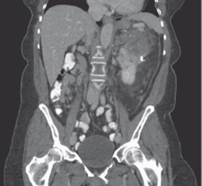
Radiologic Findings
Diagnosis
Differential Diagnosis
Discussion
Background
Clinical Findings
Related posts:


Stay updated, free articles. Join our Telegram channel

Full access? Get Clinical Tree


