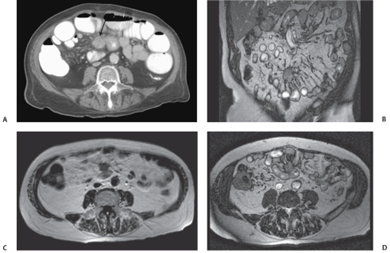
CASE 104 A 91-year-old man presents with abdominal pain and nausea. Fig. 104.1 (A) Axial CT image of the abdomen shows multiple contrast-filled, dilated small bowel loops and a small mesenteric mass (arrow) with a surrounding desmoplastic reaction and an adherent small bowel loop, which demonstrates a circumferentially thickened wall and a narrowed lumen. (B–D) MR images of the abdomen in the same patient demonstrate the small mesenteric mass that is isointense to muscle on T1 and T2; linear bands radiate from the mass and cause retraction of the adjacent small bowel mesentery. Computed tomography (CT) scan of the abdomen performed at the time of presentation demonstrates multiple dilated small bowel loops and a small mesenteric mass with a surrounding desmoplastic reaction and an adherent small bowel loop, which demonstrates a circumferentially thickened wall and a narrowed lumen. Magnetic resonance imaging (MRI) of the abdomen in the same patient demonstrates the small mesenteric mass is isointense to muscle on T1 and T2; linear bands radiate from the mass and cause retraction of the adjacent small bowel mesentery (Fig. 104.1). Small bowel carcinoid with mesenteric metastases
Clinical Presentation
Radiologic Findings
Diagnosis
Differential Diagnosis
Discussion
Background
Related posts:


Stay updated, free articles. Join our Telegram channel

Full access? Get Clinical Tree


