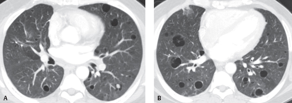
CASE 109 31-year-old woman with cough, dyspnea, and weight loss Unenhanced chest CT (lung window) (Figs. 109.1A, 109.1B) reveals bilateral, patchy ground glass opacities and thin-walled cysts that are more extensive in the lower lobes. There is mild interlobular septal thickening (Fig. 109.1B). A 1.0 cm nodule is also seen in the left lower lobe (Fig. 109.1A). Lymphocytic Interstitial Pneumonia (LIP) (Biopsy of left lower lobe nodule revealed amyloid deposition) • Nonspecific Interstitial Pneumonia (NSIP) • Hypersensitivity Pneumonitis (HP) • Various Drug Reactions • Sarcoidosis • Low-grade B-Cell Lymphoma; Mucosal-Associated Lymphoid Tissue (MALT) • Lymphangitic Carcinomatosis • Pneumocystis jiroveci Pneumonia • Mycobacterium avium-intracellulare Complex Infection • Fungal Pneumonia Fig. 109.1
 Clinical Presentation
 Radiologic Findings
 Diagnosis
 Differential Diagnosis
Immunocompetent Patients
Patients With HIV-AIDS
 Discussion
Background
Related posts:


Stay updated, free articles. Join our Telegram channel

Full access? Get Clinical Tree


