CASE 109 A middle-aged woman presents with abdominal pain and vomiting. Fig. 109.1 (A–C) Axial and coronal contrast-enhanced CT scans show dilation of the small bowel, indicative of a mechanical obstruction. The obstruction is proximal to the herniated bowel loop, seen along the anterolateral pelvic wall between the pectineal and obturator externus muscles (arrows). Axial and coronal contrast-enhanced computed tomography (CT) scans show dilation of the small bowel, indicative of a mechanical obstruction. The obstruction is proximal to the herniated bowel loop, seen along the anterolateral pelvic wall between the pectineal and obturator externus muscles (Fig. 109.1). Small bowel obstruction secondary to obturator hernia Other causes of small bowel obstruction Obturator hernias occur when the peritoneal sac and its contents protrude through the obturator canal. The hernia sac protrudes between the obturator externus and the pectineus muscle. These hernias may also be found between the superior and medial fasciculi of the obturator externus muscle or the external and internal obturator muscles.
Clinical Presentation
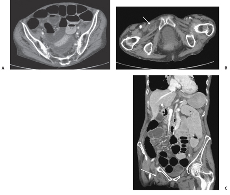
Radiologic Findings
Diagnosis
Differential Diagnosis
Discussion
Background
Clinical Findings
Related posts:


Stay updated, free articles. Join our Telegram channel

Full access? Get Clinical Tree


