Case 11
Indication: Ultrasound examination performed elsewhere raised suspicion of breast cancer.
History: Unremarkable.
Risk profile: No increased risk.
Age: 45 years.
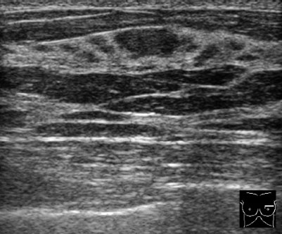
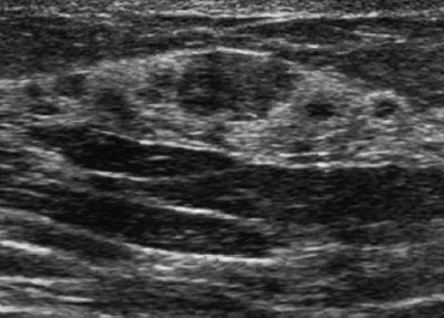
Fig. 11.1 Sonography.
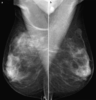
Fig. 11.2a,b Diaital mammoaraphy. MLO view.
Clinical Findings
No findings.
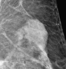
Fig. 11.3 Magnification view of the left breast, upper outer quadrant (MLO).
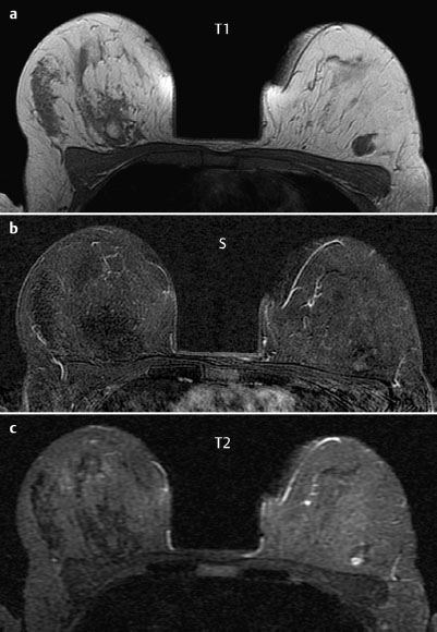
Fig. 11.4a–c Contrast-enhanced MR mammography.
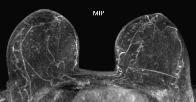
Fig. 11.5 Contrast-enhanced MR mammography. Maximum intensity projection.

|
Please characterize ultrasound, mammography, and MRI findings.
What is your preliminary diagnosis?
What are your next steps? |
Further imaging studies were performed following suspicious findings in an initial ultrasound examination.
Ultrasound
There was an oval, mostly well-defined mass with a maximum diameter of 8 mm in the upper outer quadrant of the left breast. The long axis of this lesion was parallel to the skin. Homogeneous internal structure. There were no unusual echo patterns in surrounding tissue. US BI-RADS left 3.
Mammography
Digital mammography showed bilaterally symmetric, extremely dense parenchyma, especially in the retromammary regions (ACR type 4). There were no pathological findings in the right breast. There was an oval mass with largely well-defined borders and a hypodense internal pattern in the upper outer quadrant of the left breast. Mammograms showed no calcifications and no architectural distortion. BI-RADS right 1/left 3. PGMI: G (inframammary and axillary folds visible).
MR Mammography
MRI demonstrated an oval mass in the upper outer quadrant of the left breast, consistent with ultrasound and mammographic findings. This mass showed a well-defined margin and no enhancement after administration of contrast medium. In the water-sensitive inversion recovery sequence, this lesion showed a moderate signal increase.
MRI Artifact Category: 1
MRI Density Type: 2
 Preliminary Diagnosis