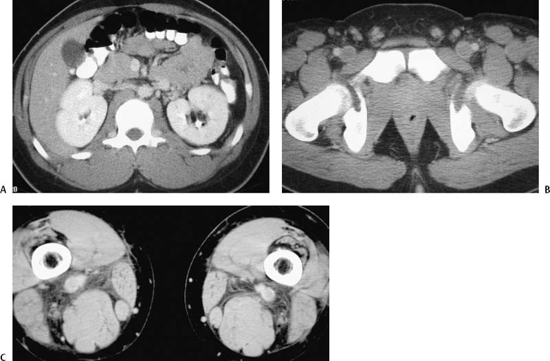
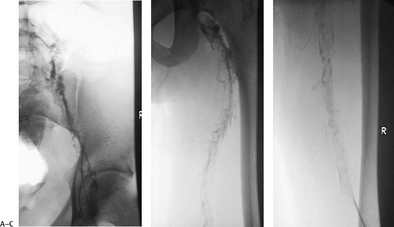
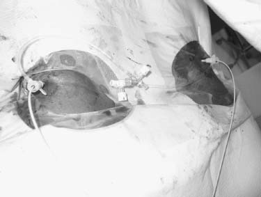
CASE 11 An 18-year-old male with no significant past medical history presented with acute bilateral leg pain and swelling. Figure 11-1 Absence of the infrarenal inferior vena cava with lower extremity thrombosis. (A) CT image shows absence of the inferior vena cava. (B) CT image shows bilateral common femoral vein thrombosis. (C) CT image shows bilateral popliteal vein thrombosis. An ultrasound exam of the legs revealed bilateral thigh and calf deep venous thromboses (DVT) extending into the pelvis. A CT exam was performed and showed absence of the inferior vena cava and extension of clot throughout both common and external iliac veins (Fig. 11-1). Additional findings included bilateral common iliac vein aneurysms and a dilated paraspinal venous plexus. Pelvic and leg deep venous thrombosis (DVT) associated with absence of the inferior vena cava. Because of the patient’s young age and active lifestyle, thrombolytic therapy was offered to potentially avert the long-term venous insufficiency associated with chronic DVT. Both thrombosed popliteal veins were punctured using sonographic guidance, and guidewires were advanced through soft thrombus into the iliac veins using fluoroscopic guidance. Venography confirmed complete thromboses of both deep venous systems and absence of prograde blood flow (Fig. 11-2). Infusion catheters were inserted into the popliteal veins and thrombolytic therapy was initiated using tissue plasminogen activator (Alteplase, Genentech, South San Francisco, California) infused at a rate of 0.25 mg per hour into each leg (Fig. 11-3). Intravenous heparin was also administered into a peripheral vein at a rate of 100 U per hour. Figure 11-3 Photograph shows bilateral popliteal sheaths inserted to perform thrombolysis.
Clinical Presentation
Radiologic Studies
Diagnosis
Treatment
Related posts:


Stay updated, free articles. Join our Telegram channel

Full access? Get Clinical Tree


