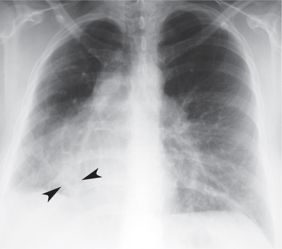
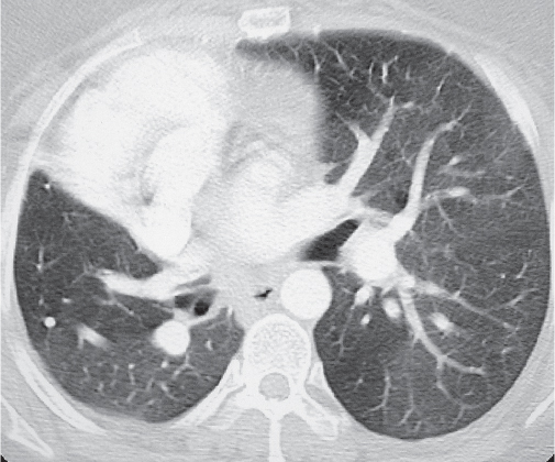
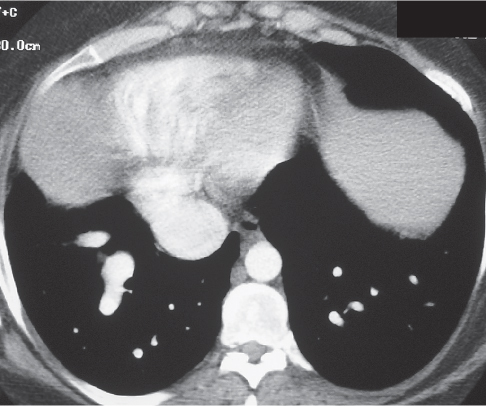
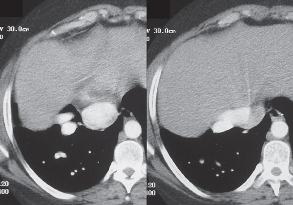
CASE 11 Asymptomatic 29-year-old woman PA (Fig. 11.1) chest X-ray demonstrates a small right lung with ipsilateral mediastinal shift and arcuate tubular opacities that course toward the right cardiophrenic angle. Note that the right bronchus is hyparterial (Fig. 11.1). Contrast-enhanced chest CT (lung window) (Fig. 11.2) demonstrates the small volume of the bilobed right lung. Contrast-enhanced chest CT (mediastinal window) (Figs. 11.3, 11.4) demonstrates paired anomalous pulmonary veins (Fig. 11.3) that drain into the inferior vena cava (Fig. 11.4). Scimitar Syndrome • Arteriovenous Malformation • Pulmonary Varix • Intralobar Sequestration • Mucoid Impaction/Bronchial Atresia Fig. 11.1 Fig. 11.2 Fig. 11.3 Fig. 11.4 Scimitar syndrome is a complex disorder also known as pulmonary venolobar syndrome and hypogenetic lung syndrome. It characteristically affects the right lung and manifests with a variety of cardiopulmonary anomalies. The term scimitar
 Clinical Presentation
 Radiologic Findings
 Diagnosis
 Differential Diagnosis
 Discussion
Background
Related posts:
![]()
Stay updated, free articles. Join our Telegram channel

Full access? Get Clinical Tree


Radiology Key
Fastest Radiology Insight Engine


