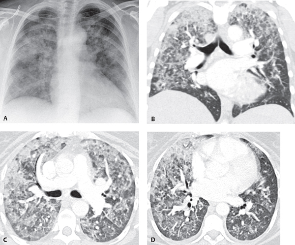
CASE 111 34-year-old man with recent sore throat, chills, and fever, subsequently developed hematuria, dysuria, cough, dyspnea, hemoptysis, and respiratory failure AP chest radiograph (Fig. 111.1A) reveals diffuse bilateral air space opacities. Chest CT (lung window), coronal reformatted image (Fig. 111.1B), and axial images (Figs. 111.1C, 111.1D) reveal diffuse bilateral ground glass opacity and patchy areas of consolidation. Diffuse Alveolar Hemorrhage; Goodpasture Syndrome Fig. 111.1 • Idiopathic Pulmonary Hemorrhage • Other Diffuse Pulmonary Hemorrhage Syndromes • Sequela of Drug Therapy • Crack Cocaine Abuse • Environmental Exposures • Bone Marrow and Heart-Lung Transplantation • Dieulafoy Disease (e.g., endobronchial vascular malformation)
 Clinical Presentation
 Radiologic Findings
 Diagnosis
 Differential Diagnosis
 Wegener Granulomatosis
 Henoch-Schonlein Purpura
 Microscopic Polyangiitis Pauci–Immune Glomerulonephritis
 Systemic Lupus Erythematosus
 Drug-Induced Coagulopathy
 Penicillamine; Nitrofurantoin; Amiodarone
 Paraquat (Zeneca Ag Products, Wilmington, DE)
 Pesticides
 Leather Conditioners
 Isocyanates
 Discussion
Background
Related posts:


Stay updated, free articles. Join our Telegram channel

Full access? Get Clinical Tree


