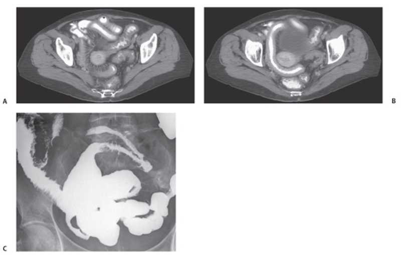
CASE 112 A 55-year-old woman presents with a 2-month history of abdominal discomfort, anorexia, and frequent bowel movements after being treated for rectal carcinoma. Fig. 112.1 (A,B) Axial images from a contrast CT scan show thickening pelvic small bowel loops with loss of normal folds. (C) Small bowel barium series demonstrates deep ulcers in the small bowel, mucosal edema, and strictures. Axial images from a contrast-enhanced computed tomography (CT) scan show thickening pelvic small bowel loops with loss of normal folds. A small bowel barium series demonstrates deep ulcers in the small bowel, mucosal edema, and strictures (Fig. 112.1). Radiation enteritis
Clinical Presentation
Radiologic Findings
Diagnosis
Differential Diagnosis
Discussion
Related posts:


Stay updated, free articles. Join our Telegram channel

Full access? Get Clinical Tree


