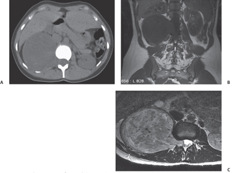
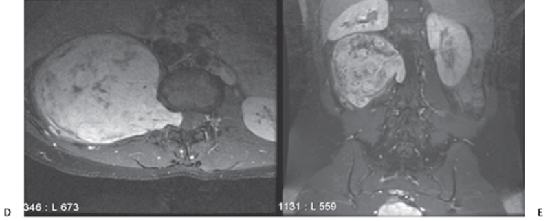
CASE 113 A 35-year-old woman with a past history of Hodgkin lymphoma presents with right-sided abdominal pain. Fig. 113.1 (A) A noncontrast CT scan shows a large, asymmetrically dumbbell-shaped mass arising from a right neural foramen at a superior lumbar level. The mass measures soft tissue attenuation and causes widening of the right neural foramen. (B–E) On MRI, the mass is homogeneously hypointense to muscle on T1, predominantly hyperintense on T2, and demonstrates intense, heterogeneous enhancement after the administration of gadolinium. Coronal imaging confirms that the mass arises from a neural foramen and superiorly displaces the right kidney. A noncontrast computed tomography (CT) scan shows a large, asymmetrically dumbbell-shaped mass arising from a right neural foramen at a superior lumbar level. The mass measures soft tissue attenuation and causes widening of the right neural foramen. On magnetic resonance imaging (MRI), the mass is homogeneously hypointense to muscle on T1, predominantly hyperintense on T2, and demonstrates intense, heterogeneous enhancement after the administration of gadolinium. Coronal imaging confirms that the mass arises from a neural foramen and superiorly displaces the right kidney (Fig. 113.1). Retroperitoneal schwannoma (also known as a neurilemoma or neurinoma)
Clinical Presentation


Radiologic Findings
Diagnosis
Differential Diagnosis
Stay updated, free articles. Join our Telegram channel

Full access? Get Clinical Tree


