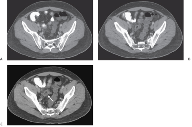
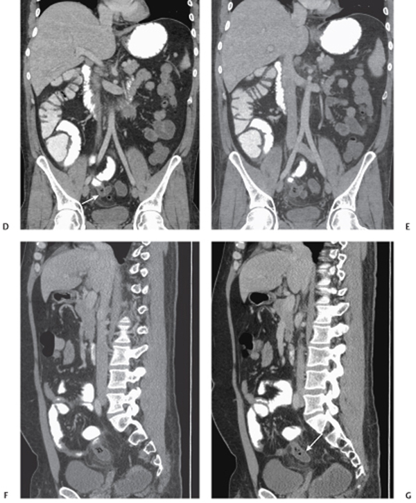
CASE 115 A 30-year-old man presents with a 1-day history of emesis and diarrhea. Fig. 115.1 (A–C) Axial, (D,E) coronal, and (F,G) sagittal images of the abdomen and pelvis show a tubular structure arising from the distal small bowel (arrows) filled with gas and particulate material. There is accompanying soft tissue stranding seen surrounding this structure. Axial, coronal, and sagittal images of the abdomen and pelvis show a tubular structure arising from the distal small bowel filled with gas and particulate material. There is accompanying soft tissue stranding seen surrounding this structure (Fig. 115.1). Perforated Meckel diverticulum
Clinical Presentation
Radiologic Findings
Diagnosis
Differential Diagnosis
Discussion
Background
Related posts:


Stay updated, free articles. Join our Telegram channel

Full access? Get Clinical Tree


