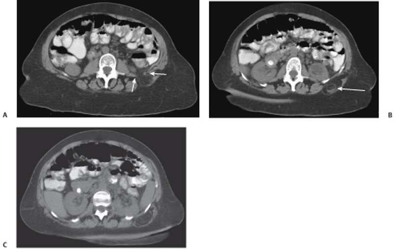
CASE 119 A 64-year-old woman presents with vague abdominal pain. Fig. 119.1 (A) Axial contrast-enhanced CT image shows retroperitoneal fat and stranding passing through an abdominal wall defect (arrows) in the left flank. (B,C) There is soft tissue density from fat necrosis (arrow) seen within the herniated fat. Axial contrast-enhanced computed tomography (CT) images show retroperitoneal fat and stranding passing through an abdominal wall defect in the left flank. There is soft tissue density seen within the herniated fat (Fig. 119.1). Lumbar hernia of Grynfeltt through the superior lumbar triangle
Clinical Presentation
Radiologic Findings
Diagnosis
Differential Diagnosis
Discussion
Background
Related posts:


Stay updated, free articles. Join our Telegram channel

Full access? Get Clinical Tree


