Case 12
Indication: Screening.
History: Unremarkable.
Risk profile: No increased risk.
Age: 48 years.
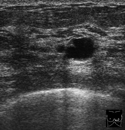
Fig. 12.1 Sonography.
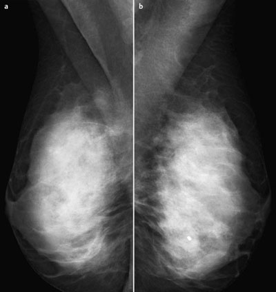
Fig. 12.2a,b Digital mammography, MLO view.
Clinical Findings
Normal.
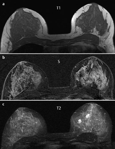
Fig. 12.3a–c Contrast-enhanced MR mammography.
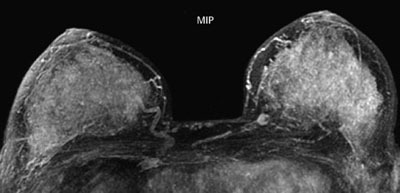
Fig. 12.4 Contrast-enhanced MR mammography. Maximum intensity projection.

|
Please characterize ultrasound, mammography, and MRI findings.
What is your preliminary diagnosis?
What are your next steps? |
Because of the density of the parenchyma, mammography was performed in one view only (MLO) and supplemented by contrast-enhanced MR mammography (so-called optipack-concept).
Ultrasound
The US image is of the lower inner quadrant of the left breast. It demonstrates a cyst compatible with the information provided by T2-weighted MRI. US BI-RADS left 2.
Mammography
The mammogram shows extremely dense parenchyma of both breasts, ACR type 4. Under these limiting conditions, no abnormalities were detectable. The macrocalcifications in the center of the left breast can be considered harmless and irrelevant. (BI-RADS right 1/left 1). PGMI: MLO view G (inframammary fold not clearly depicted).
MR Mammography
Subtraction images were obtained by subtracting the precontrast image from images derived from the second measurement after administration of contrast (approx. 3 minutes after introduction of contrast into peripheral veins). These early subtraction images show strong enhancement of all glandular tissue, meaning that It no further differentiation of circumscribed lesions was possible, However, a round enhancing area was observed in the inner region of the left breast (not shown on the slices reproduced here).
MRI Artifact Category: 2
MRI Density Type: 4
 Preliminary Diagnosis


