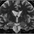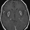
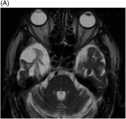
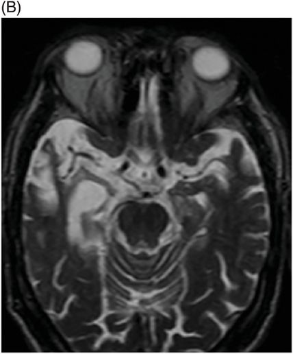
(A–B) Axial T2WI through the temporal lobes demonstrates striking atrophy of the right anterior and medial temporal lobe.
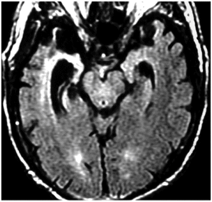
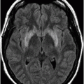
Axial FLAIR image through the temporal lobes.
Right Temporal Variant of Frontotemporal Lobar Degeneration
Primary Diagnosis
Right temporal variant of frontotemporal lobar degeneration
Imaging Findings
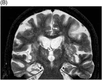
Fig. 12.1: (A) Coronal T2WI through the anterior temporal lobe demonstrated striking atrophy of the right anterior and inferior temporal lobe. Atrophy of the frontal and left temporal lobes is present; however, it is mild, in comparison to the right temporal lobe. (B) Coronal T2WI obtained more posteriorly, demonstrated striking atrophy of the right medial temporal lobe, in comparison to the remainder of the visualized areas of brain. Fig. 12.2: (A–B) Axial T2WI through the temporal lobes demonstrates striking atrophy of the right anterior and medial temporal lobe. Fig. 12.3: Axial FLAIR image through the temporal lobes demonstrated increased FLAIR signal in the remainder of the anterior and medial temporal lobe, in addition to the severe atrophy of the right anterior and medial temporal lobe.
Stay updated, free articles. Join our Telegram channel

Full access? Get Clinical Tree


