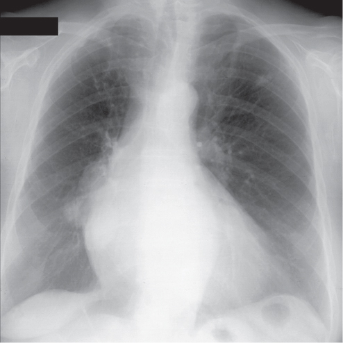
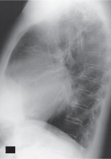
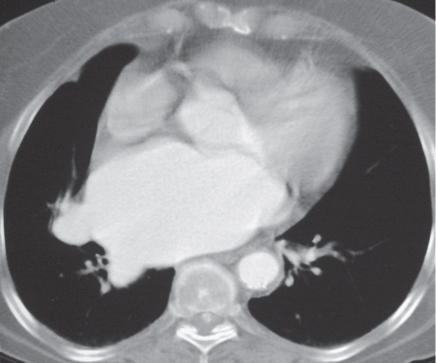
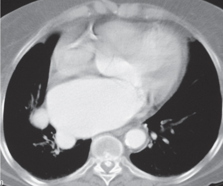
CASE 12 65-year-old woman with chest pain PA (Fig. 12.1) and lateral (Fig. 12.2) chest radiographs demonstrate a multi-lobular soft-tissue mass that projects over the right hilum (Fig. 12.1) and focal enlargement of the superior aspect of the left atrium (Fig. 12.2). Nodular upper lobe parenchymal opacities represented remote granulomatous infection. Contrast-enhanced chest CT (Figs. 12.3, 12.4) shows that the lobular opacities seen on radiography correspond to focal dilatation of the pulmonary veins as they enter an enlarged left atrium. Pulmonary Varix • Indeterminate Pulmonary Nodule/Mass Fig. 12.1 Fig. 12.2 Fig. 12.3 Fig. 12.4 Pulmonary varix is a rare lesion characterized by focal non-obstructive aneurysmal enlargement of one or more pulmonary veins prior to entering the left atrium. Both acquired and congenital etiologies of pulmonary varix have been proposed. Pulmonary varix is typically associated with mitral valve dysfunction, particularly insufficiency. In these cases, the right pulmonary veins are typically affected.
 Clinical Presentation
 Radiologic Findings
 Diagnosis
 Differential Diagnosis


 Discussion
Background
Etiology
Clinical Findings
![]()
Stay updated, free articles. Join our Telegram channel

Full access? Get Clinical Tree


Radiology Key
Fastest Radiology Insight Engine


