VIII Bladder
CASE 120
Clinical Presentation
A 65-year-old man presents with suprapubic pain.
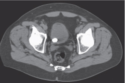
Fig. 120.1 Noncontrast axial CT image in the supine position demonstrates a calcified round stone in the dependent location of the bladder.
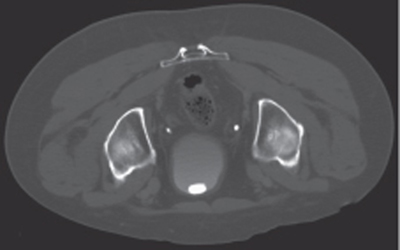
Fig. 120.2 Postcontrast image in the same patient obtained in the prone position demonstrates a change in the position of this stone, which is freely mobile within the bladder lumen.
Radiologic Findings
Noncontrast axial computed tomography (CT) image in the supine position demonstrates a calcified round stone in the dependent portion of the bladder (Fig. 120.1). Additional postcontrast imaging obtained in the prone position demonstrates a change in the position of this stone, which is freely mobile within the bladder lumen (Fig. 120.2).
Diagnosis
Bladder stone
Differential Diagnosis
- Bladder tumor
- Blood clot
Discussion
Background
Related posts:


Stay updated, free articles. Join our Telegram channel

Full access? Get Clinical Tree


