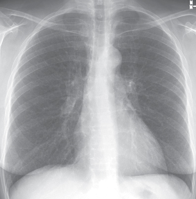
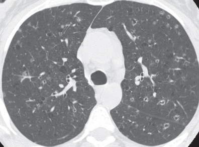
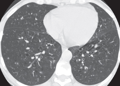
CASE 120 38-year-old woman smoker with cough and dyspnea PA chest radiograph (Fig. 120.1) demonstrates normal lung volume without visible pulmonary abnormalities. HRCT (Figs. 120.2, 120.3) shows subtle bilateral upper lobe–predominant small pulmonary cysts with nodular irregular cyst walls and scattered small nodules with ill-defined borders (Fig. 120.2). Note the relative sparing of the lung bases (Fig. 120.3). Pulmonary Langerhans’ Cell Histiocytosis • Infection • Emphysema • Bronchiectasis • Lymphangioleiomyomatosis Pulmonary Langerhans’ cell histiocytosis (PLCH) is a chronic, progressive interstitial lung disease that results from abnormal non-malignant proliferation of monoclonal Langerhans’ cells. Multiple organs may be affected, including bone, pituitary gland, mucous membranes, skin, lymph nodes, and liver. Fig. 120.1 Fig. 120.2 Fig. 120.3
 Clinical Presentation
 Radiologic Findings
 Diagnosis
 Differential Diagnosis
 Discussion
Background
Etiology
Related posts:
![]()
Stay updated, free articles. Join our Telegram channel

Full access? Get Clinical Tree


Radiology Key
Fastest Radiology Insight Engine


