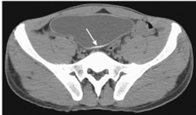
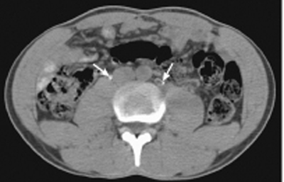
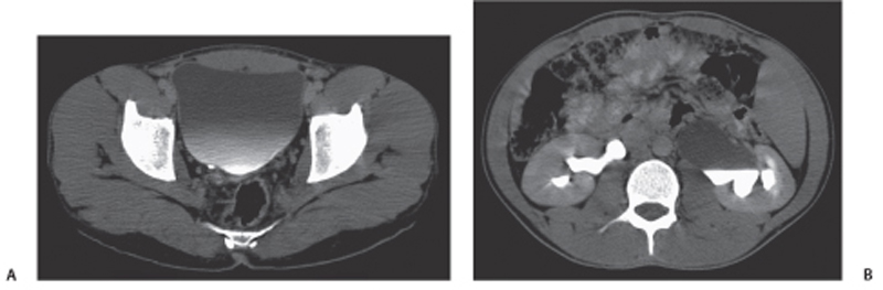
CASE 122 A 53-year-old man, an immigrant from the Middle East, presents with hematuria. Fig. 122.1 Noncontrast image through the pelvis demonstrates diffuse circumferential calcification (arrow) in the wall of the bladder. Fig. 122.2 Noncontrast image through the lower abdomen demonstrates circumferential calcification (arrows) in the wall of the ureters. Fig. 122.3 (A) The circumferential wall calcification is difficult to see following contrast administration. (B) There is accompanying mild dilatation of the ureters and left hydronephrosis. Noncontrast images through the pelvis demonstrate diffuse circumferential calcification in the wall of the bladder and both ureters (Figs. 122.1 and 122.2). There is mild dilatation of the ureters and left hydronephrosis (Fig. 122.3
Clinical Presentation
Radiologic Findings
![]()
Stay updated, free articles. Join our Telegram channel


Full access? Get Clinical Tree


