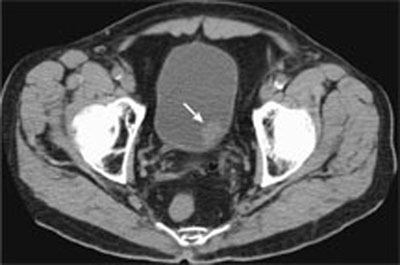
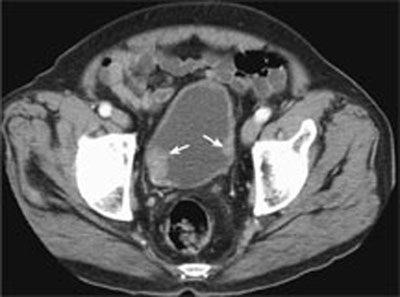
CASE 125 A 59-year-old man presents with painless gross hematuria. Fig. 125.1 Noncontrast CT image through the pelvis shows a polypoid lesion (arrow) arising from the left posterior wall of the bladder. Noncontrast computed tomography (CT) image through the pelvis shows a polypoid lesion arising from the left posterior wall of the bladder (Fig. 125.1). Transitional cell carcinoma of the bladder Bladder cancer is ranked as the fourth most common malignancy in the United States. Transitional cell carcinoma accounts for 90% of cases and has a propensity to be multicentric with synchronous and metachronous bladder and upper tract tumors. Multicentric bladder tumors occur in 30 to 40% of cases (Fig. 125.2). Upper tract tumors occur in 3 to 5% of bladder tumor cases and are seen most frequently when multiple bladder lesions are present. Fig. 125.2 Contrast-enhanced CT image of the pelvis shows multiple enhancing lesions (arrows) arising from the lateral bladder wall.
Clinical Presentation
Radiologic Findings
Diagnosis
Differential Diagnosis
Discussion
Background
Clinical Findings
Related posts:


Stay updated, free articles. Join our Telegram channel

Full access? Get Clinical Tree


