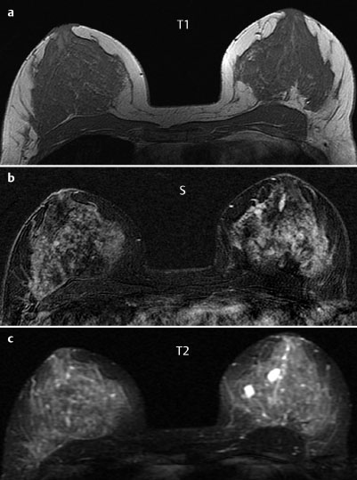
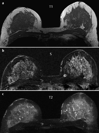
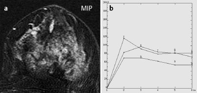
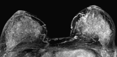
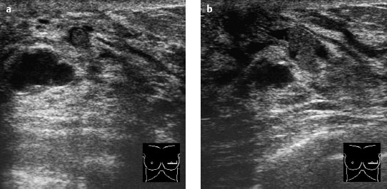
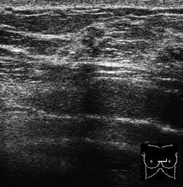
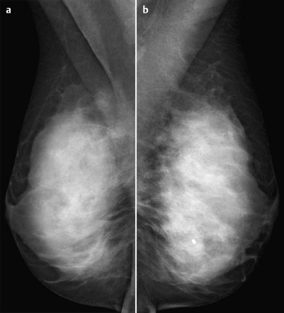
Case 13 Indication: Screening. History: Unremarkable. Risk profile: Not increased. Age: 48 years. In addition to the conventional MR mammography imaging seen in Case 12, a very early subtraction (images of first measurement after contrast minus precontrast images) was performed. Fig. 13.1 a-c MR mammography (earliest subtraction). Fig. 13.2a–c MR mammography (earliest subtraction). Fig. 13.3a,b Signal-to-time curves of the linear enhancement. Fig. 13.4a,b Signal-to-time curves of the round enhancement. Fig. 13.5a,b Digital mammography, MLO view. Normal. Fig. 13.6 Contrast-enhanced MR mammography. Maximum intensity projection. Fig. 13.7a,b Targeted sonography in region of MRI findings, behind the left nipple. Fig. 13.8 Targeted sonography in region of MRI findings, left upper inner quadrant. Please characterize ultrasound, mammography, and MRI findings. What is your preliminary diagnosis? What are your next steps?

Clinical Findings


Stay updated, free articles. Join our Telegram channel

Full access? Get Clinical Tree


