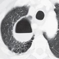
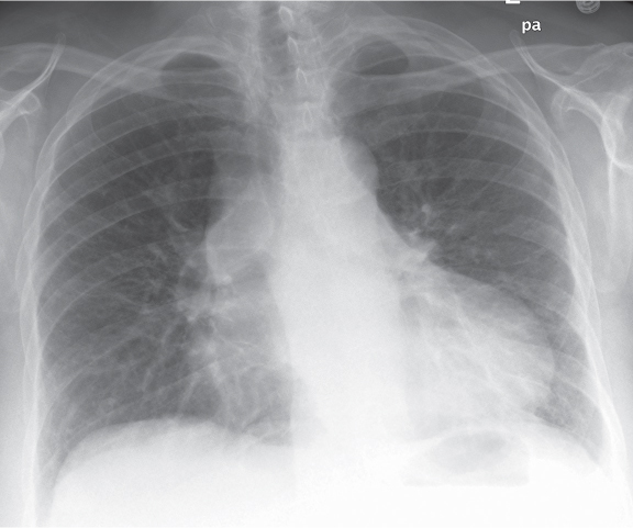
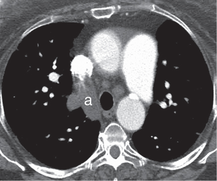
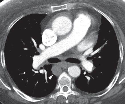
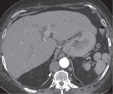
CASE 13 68-year-old woman admitted for pacemaker placement PA chest radiograph (Fig. 13.1) demonstrates cardiomegaly, marked enlargement of the azygos arch and vein, and bilateral hyparterial bronchi. Contrast-enhanced CT (mediastinal window) (Figs. 13.2, 13.3, 13.4) demonstrates bilateral hyparterial bronchi (Fig. 13.3), enlargement of the azygos arch (a) (Fig. 13.2), and descending azygos vein (arrowhead) (Figs. 13.3, 13.4), consistent with azygos continuation of the inferior vena cava and multiple left-sided spleens (polysplenia) (Fig. 13.4). Heterotaxy Syndrome: Polysplenia, Azygos Continuation of the Inferior Vena Cava • Azygos Continuation of the Inferior Vena Cava with Situs Solitus Fig. 13.1 Fig. 13.2 Fig. 13.3 Fig. 13.4 Visceroatrial situs (location, position) refers to the location of the atria with respect to the midline and to other organs. The majority of the population exhibit normal visceroatrial situs (situs solitus) characterized by a morphologic right (systemic) atrium on the right side and a contralateral morphologic left (pulmonary) atrium. The pulmonary anatomy is characterized by a tri-lobed right lung with ipsilateral liver, gallbladder, and inferior vena cava and a bi-lobed left lung with ipsilateral stomach, single spleen, and aortic arch. The incidence of congenital heart disease in patients with situs solitus is approximately 0.8%. Approximately 0.1% of the population exhibit situs inversus, characterized by mirror image location of the atria, major vessels, and abdominal structures. These patients have a 3–5% incidence of congenital heart disease. The terms heterotaxy, isomerism, and situs ambiguous
 Clinical Presentation
 Radiologic Findings
 Diagnosis
 Differential Diagnosis
 Discussion
Background
![]()
Stay updated, free articles. Join our Telegram channel

Full access? Get Clinical Tree


Radiology Key
Fastest Radiology Insight Engine

