CASE 13 A 2-week-old infant presents with facial deformity. Figure 13A Three-dimensional CT demonstrates bilateral midfacial hypoplasia. There is hypoplasia of the malar bone, with absence of the zygomatic arch and hypoplasia of the mandibular ramus and condyle. The temporomandibular joint is absent (Fig. 13A1). The temporal bone is not fully developed, and the external auditory canal is abnormally located inferiorly. Soft tissue imaging demonstrates an abnormal pinna (Fig. 13A2). These abnormalities were essentially symmetrical. Lateral facial dysplasias: Treacher Collins syndrome (TCS), also called mandibulofacial dysostosis (MFD1) or Treacher Collins-Franceschetti syndrome (TCOF) TCS is a disorder of craniofacial development. An autosomal dominant syndrome, it is found in 1 per 50,000 live births. The TCOF1 mutation is found in 78% of patients. Familial in 40% of cases, it arises as new spontaneous mutation in 60%. The obligatory features of TCOF are marked hypoplasia of the malar bone, hypoplasia of the mandibular ramus and condyle, obliteration of the frontonasal angle, colobomas of the lower eyelids, and malformation of the eyelashes. Various explanations have been offered regarding the pathogenesis of TCS: problems of differentiation of the branchial arch mesoderm preventing normal facial bone development, abnormal ossification of the viscerocranium, even ischemia from stapedial artery hypoplasia or defect of ectomesenchymal cells within the developing trigeminal ganglia.
Clinical Presentation
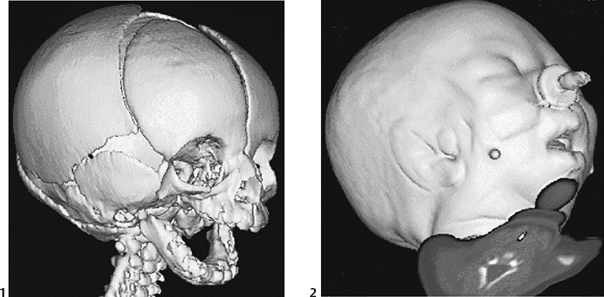
Radiologic Findings
Diagnosis
Differential Diagnosis
Discussion
Background
Etiology
Genetics
Related posts:


Stay updated, free articles. Join our Telegram channel

Full access? Get Clinical Tree


