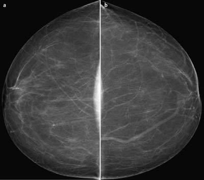
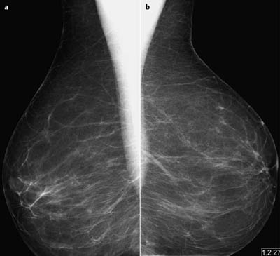
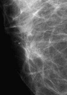
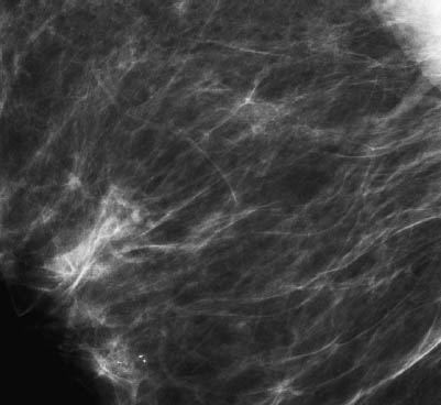
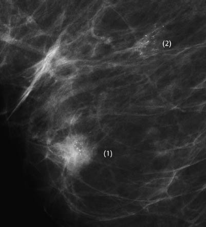
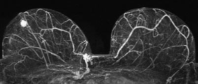
Case 14 Indication: Screening. History: Unremarkable. Risk profile: No increased risk. Age: 57 years. Fig. 14.1 a,b Digital mammography, CC view [imaging not performed by authors]. Fig. 14.2a,b Digital mammography, MLO view [imaging not performed by authors]. Normal. No suspicious findings (not shown). Fig. 14.3 Magnification view of the right breast, behind the nipple. Fig. 14.4 Magnification view at first examination. Fig. 14.5 Magnification view at follow-up 7 months later. Please characterize the calcifications on the mammograms. What is your preliminary diagnosis? What are your next steps? Mammography showed predominantly lipomatous tissue in both breasts, ACR type 1. There were no lesions or masses. There is, however, a cluster of polymorphous microcalcifications in the region behind the nipple of the right breast (Fig. 14.4). These calcifications were categorized as BI-RADS 3. (BI-RADS right 3/left 1). Follow-up mammography of the right breast in 6 months was recommended. The follow-up mammography 7 months later (Fig. 14.5), showed no changes and the findings were again categorized as BI-RADS 3. Another follow-up mammography was recommended in a further 6 months. Unfortunately, the patient did not attend for the follow-up mammography 6 months later, but rather 24 months after the original examination (Fig. 14.6). There was a round lesion containing microcalcifications behind the nipple of the right breast-i.e., exactly where calcifications had been identified 2 years previously (1). There was a further cluster of polymorphous microcalcifications in the central part of the right breast (2). Retrospectively, this second cluster was not visible in the earlier images. MRI demonstrated a hypervascularized mass behind the right nipple, corresponding to the lesion with calcifications (1) as seen in mammography (Fig. 14.6). There was no increased vascularization corresponding to the second cluster of microcalcifications (2) in the center of the right breast. Fig. 14.6 Mammography 24 months after first examination. Fig. 14.7 MR imaging performed at the same time as the mammogram seen in Fig. 14.6.
Clinical Findings
Ultrasound

Mammography
Mammography (Fig. 14.6)
MR Mammography
BI-RADS Categorization (First examination and follow-up after 7 months) | ||
Clinical Findings | right 1 | left 1 |
Ultrasound | right 1 | left 1 |
Mammography | right 3 | left 1 |
MR Mammography | – | – |
BI-RADS Total | right 3 | left 1 |
 Preliminary Diagnosis
Bifocal breast cancer of the right breast (presumed invasive in the region behind the nipple, presumed intraductal in the central region).
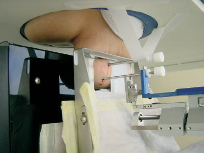
Fig. 14.8 Simultaneous stereotactic vacuum biopsy of both findings.
BI-RADS Categorization (Follow-up examination after 2 years) | ||
Clinical Findings | right 1 | left 1 |
Ultrasound | right 1 | left 1 |
Mammography | right 5(bifocal) | left 1 |
MR Mammography | right 5 | left 1 |
BI-RADS Total | right 5 | left 1 |
Procedure
Stereotactic vacuum biopsy.
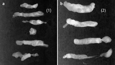
Fig. 14.9 Specimen radiograms.
a Specimen from the lesion behind the nipple (marked [1] in Fig. 14.6).
b Specimen from the central clustered calcifications (marked [2] in Fig. 14.6).
Diagnosis
Right breast behind nipple: IDC pT1 b (5 mm), pNO (0/15), R0, G2
Right breast central: DCIS (no residual tumor in excised specimens)


Stay updated, free articles. Join our Telegram channel

Full access? Get Clinical Tree


