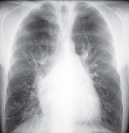
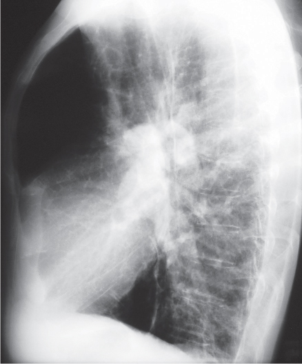
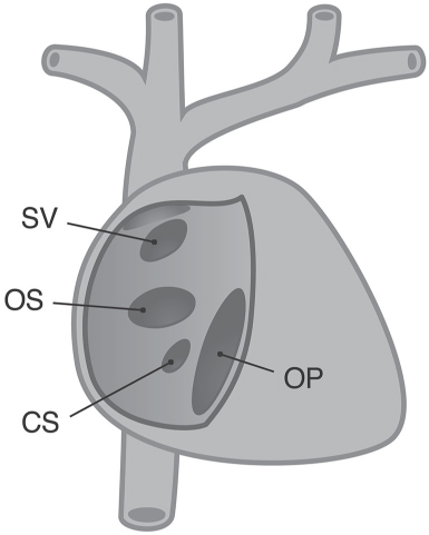
CASE 14 35-year-old man evaluated for a murmur found on a routine physical examination PA (Fig. 14.1) and lateral (Fig. 14.2) chest radiographs demonstrate cardiomegaly and over-circulation. Note the enlarged central and peripheral pulmonary arteries. Atrial Septal Defect • Other Left-to-Right Shunts; Ventricular Septal Defect; Patent Ductus Arteriosus • Pulmonary Arterial Hypertension Fig. 14.1 Fig. 14.2 Atrial septal defect (ASD) accounts for approximately 10% of congenital heart disease and is a common congenital cardiac lesion among those that initially manifest in adulthood. It represents approximately 30% of all congenital cardiac lesions in patients over the age of 40 years. ASD may be an isolated condition or may be associated with other cardiovascular anomalies. Atrial septal defect represents a congenital defect in the interatrial septum that results in communication between the two atria. ASD is classified based on anatomic location (Fig. 14.3). Ostium secundum ASD is the most common type, located at the fossa ovalis; ostium primum ASD is rare but represents the second most common ASD, is located in the inferior atrial septum, and is associated with partial atrioventricular septal defect (including a cleft mitral valve); sinus venosus ASD is rare, is located at the superior aspect of the septum near the orifice of the superior vena cava, and is associated with anomalous pulmonary venous drainage in up to 90% of cases; coronary sinus ASD represents unroofing of the coronary sinus with resultant interatrial communication and is associated with persistent left superior vena cava, partial anomalous pulmonary venous connections, and mitral valve prolapse. Patients with ostium secundum ASD may remain asymptomatic until the second or third decade of life. Women are more commonly affected than men, with a 3:2 female-to-male ratio. Some patients are diagnosed incidentally because of a left sternal murmur, which is typically systolic with a split fixed second heart sound. Approximately half of patients over the age of 40 years are symptomatic and present with exertional dyspnea, fatigue, chest pain from atrial flutter/fibrillation, fever from infective endocarditis, paradoxical emboli, clinical right heart failure, and/or pulmonary hypertension. Palpitations and recurrent pulmonary infections are also reported. Approximately 15% of affected patients also have mitral insufficiency. ASD is described as part of the autosomal dominant Holt-Oram syndrome Fig. 14.3
 Clinical Presentation
 Radiologic Findings
 Diagnosis
 Differential Diagnosis
 Discussion
Background
Etiology
Clinical Findings
Related posts:
![]()
Stay updated, free articles. Join our Telegram channel

Full access? Get Clinical Tree


Radiology Key
Fastest Radiology Insight Engine


