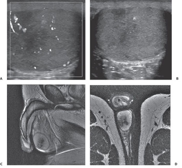
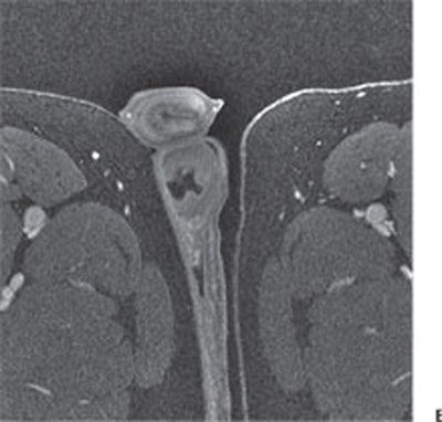
CASE 144 A 26-year-old man complains of dull pain in the right scrotum. Fig. 144.1 (A,B) Ultrasound images of the right testicle show an ill-defined, hypoechoic area with no increased Doppler flow. (C,D) Sagittal and axial T2-weighted images show an ill-defined, hypointense area within a hyperintense normal testis. (E) On a postgadoliniumenhanced T1-weighted image, there is a focal area that shows a lack of normal enhancement. Ultrasound of the right testis shows an ill-defined, hypoechoic area with no increase in Doppler flow. T2-weighted magnetic resonance imaging (MRI) shows an ill-defined, hypointense area in the right testis, which shows a lack of enhancement on the postgadolinium-enhanced T1-weighted image (Fig. 144.1). Segmental testicular infarct
Clinical Presentation
Radiologic Findings
Diagnosis
Differential Diagnosis
Related posts:


Stay updated, free articles. Join our Telegram channel

Full access? Get Clinical Tree


