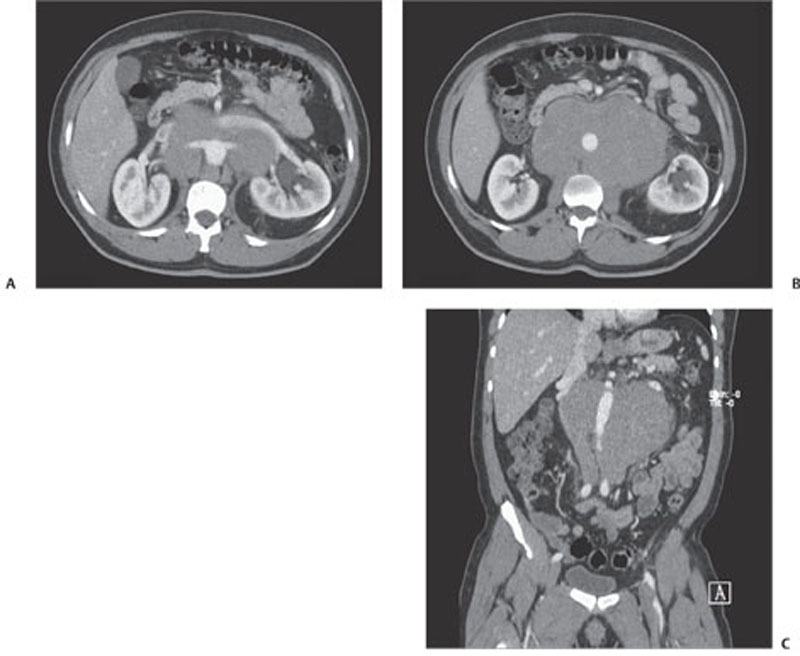
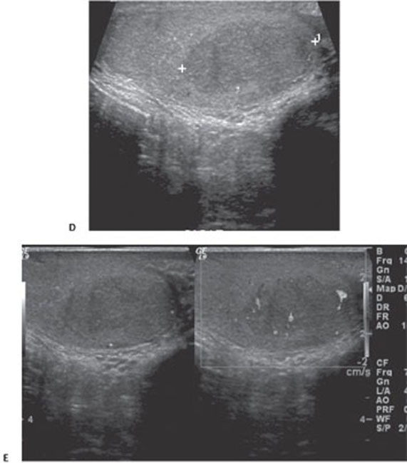
145 Left Malignant Germ Cell Tumor of the Testis with Metastatic Bulky Retroperitoneal Adenopathy

CASE 145


Clinical Presentation


A 31-year-old male patient complains of ill-defined abdominal pain.


image


image

Fig. 145.1 (A–C) Contrast-enhanced axial and coronal images show bulky retroperitoneal adenopathy. (D,E) Ultrasound images of the left testicle show a well-defined, hypoechoic seminoma with increased Doppler flow.


Radiologic Findings


Contrast-enhanced computed tomography (CT) images (Fig. 145.1) show bulky retroperitoneal adenopathy centered at the level of the renal hila. Subsequent ultrasound images of the left testis shows a well-defined, hypoechoic, intratesticular mass with increased Doppler flow.


Diagnosis


Left malignant germ cell tumor of the testis with metastatic bulky retroperitoneal adenopathy


Differential Diagnosis


  • Testicular hematoma


  • Testicular lymphoma
  • Focal segmental infarct
  • Epidermoid cyst

Stay updated, free articles. Join our Telegram channel

Dec 26, 2015 | Posted by in GASTROINTESTINAL IMAGING | Comments Off on 145 Left Malignant Germ Cell Tumor of the Testis with Metastatic Bulky Retroperitoneal Adenopathy

Full access? Get Clinical Tree

Get Clinical Tree app for offline access