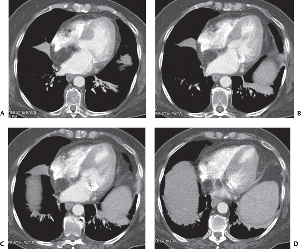
CASE 145 89-year-old woman with shortness of breath Contrast-enhanced chest CT (Figs. 145.1A, 145.1B, 145.1C, 145.1D) (mediastinal window) through the four chambers of the heart reveals a smooth-bordered fatty attenuation mass within the interatrial septum that straddles the fossa ovalis, resulting in a dumbbell shape. Note the mild deformity of the right and left atrial walls that form a smooth interface with the mass, and the absence of an intracavitary component. Right middle lobe atelectasis, left lower lobe air space disease with associated bronchiectasis, and mitral valve annulus calcification are also present. Fig. 145.1 Lipomatous Hyperplasia of the Interatrial Septum • Myxoma • Fibroma • Fibroelastoma • Lipoma • Liposarcoma Lipomatous hyperplasia of the interatrial septum (LHAS) represents benign adipose cell hyperplasia within the interatrial septum of the heart. The prevalence of LHAS is estimated to be 1–8%. Embryologically, the interatrial septum is formed by fusion of the septum primum and the septum secundum. These early outgrowths of tissue from the walls of the immature atria fuse following birth, forming the interatrial septum. In some cases, mesenchymal cells carried along with this immature tissue become trapped within the interatrial septum during the fusion process. These mesenchymal cells later develop into adipocytes when appropriately stimulated.
 Clinical Presentation
 Radiologic Findings

 Diagnosis
 Differential Diagnosis
Primary Benign Tumors in the Region of the Interatrial Septum
Fat-Containing Cardiac Tumors
 Discussion
Background
Etiology
Clinical Findings
Stay updated, free articles. Join our Telegram channel

Full access? Get Clinical Tree


