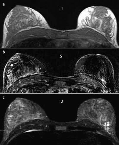
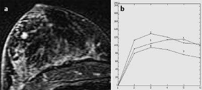

MRM score | Finding | Points |
Shape | round | 0 |
Border | ill-defined | 1 |
CM Distribution | homogeneous | 0 |
Initial Signal Intensity Increase | strong | 2 |
Post-initial Signal Intensity Character | wash-out | 2 |
MRI score (points) |
| 5 |
MRI BI-RADS |
| 4 |
 Preliminary Diagnosis
Carcinoma, fibroadenoma
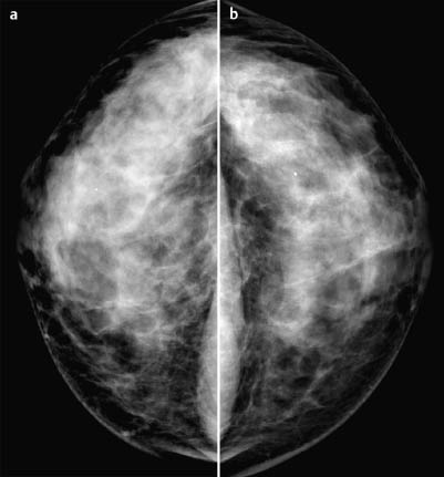
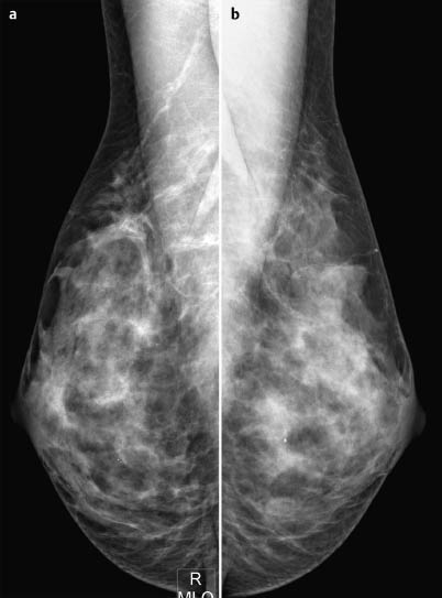
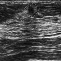
Clinical Findings | right 3 | left 1 |
Ultrasound | right 5 | left 1 |
Mammography | right 1 | left 1 |
MR Mammography | right 4 | left 1 |
BI-RADS Total | right 5 | left 1 |
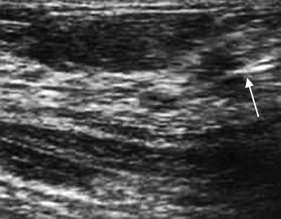
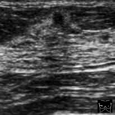
Fig. 15.8 US-guided FNAP with needle visible in lesion (arrow)
Procedure
Normally: histopathological evaluation of the palpable mass following US-guided core biopsy.
Procedure followed here (departing from guidelines)
US-guided fine-needle aspiration cytology (FNAP), because of the small size of the lesion.
Cytology
Prolific cancer cells. Category C5.
Histology
Invasive lobular carcinoma 6 mm in diameter. Axillary lymph node status normal.
ILC pT1b, pN0, M0, G2.


Stay updated, free articles. Join our Telegram channel

Full access? Get Clinical Tree


