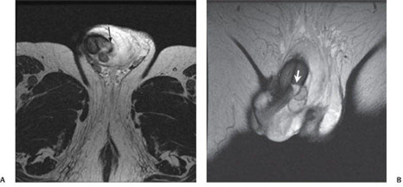
CASE 151 A 30-year-old man presents with acute, painful swelling of the penis following vaginal intercourse. Fig. 151.1 (A) Axial T2-weighted MR image shows focal discontinuity in the tunica albuginea (arrow). (B) Coronal T2-weighted MR image shows the transverse tear of the corpus cavernosum (arrow) and penile hematoma. T2-weighted magnetic resonance (MR) images show focal discontinuity in the tunica albuginea associated with a transverse tear of the corpus cavernosum and penile hematoma (Fig. 151.1). Penile fracture Imaging findings when present with a history are diagnostic of this condition.
Clinical Presentation
Radiologic Findings
Diagnosis
Differential Diagnosis
Discussion
Background
Related posts:


Stay updated, free articles. Join our Telegram channel

Full access? Get Clinical Tree


