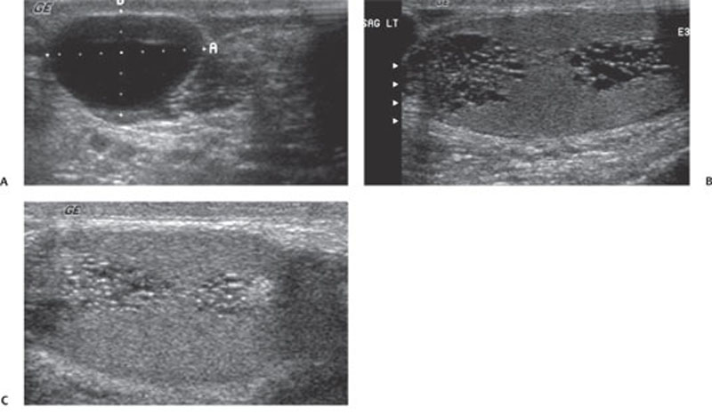
CASE 153 A 66-year-old man presents with a left scrotal mass on routine physical examination. Fig. 153.1 (A) Scrotal ultrasound reveals a 1.9 × 1.3 cm cyst within the left epididymal head likely corresponding to the palpable abnormality. Tubular and branching cystic structures are identified within the (B) left and (C) right mediastinum testes. Scrotal ultrasound reveals a 1.9 × 1.3 cm cyst within the left epididymal head, most likely corresponding to the palpable abnormality. Tubular and branching cystic structures are identified within the left and right mediastinum testes (Fig. 153.1). Tubular ectasia of the rete testes. Alternative names are dilatation, cystic dilatation, cystic ectasia, and seminiferous tubular ectasia of the rete testes.
Clinical Presentation

Radiologic Findings
Diagnosis
Differential Diagnosis
Discussion
Background
Stay updated, free articles. Join our Telegram channel

Full access? Get Clinical Tree


