CASE 154 A 58-year-old man with recently diagnosed prostate cancer and a remote history of trauma to the left side of the body following a high-speed motor vehicle accident presents for a staging computed tomography (CT) examination. Fig. 154.1 (A,B) Delayed contrast-enhanced CT scans reveal an enlarged prostate with a prominent median lobe projecting into the bladder. A lobulated soft tissue pelvic mass is seen along the left pelvic side wall (arrow). This was felt to represent a nodal mass, and patient was referred for MRI. (C) Coronal contrast-enhanced T1-weighted image reveals enlarged and tortuous enhancing vessels within the left pelvis. (D) Volume-rendered MR image demonstrates an abnormal tangle of vessels within the left pelvis supplied by an enlarged left internal iliac artery. Delayed contrast-enhanced CT scans (Fig. 154.1A,B) reveal an enlarged prostate with a prominent median lobe projecting into the bladder. A lobulated soft tissue pelvic mass is seen along the left pelvic side wall. This was felt to represent a nodal mass, and the patient was referred for magnetic resonance imaging (MRI). Coronal contrast-enhanced T1-weighted image (Fig. 154.1C) reveals enlarged and tortuous enhancing vessels within the left pelvis. Volume-rendered MR image (Fig. 154.1D) demonstrates an abnormal tangle of vessels within the left pelvis supplied by an enlarged left internal iliac artery.
Clinical Presentation
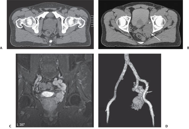
Radiologic Findings
Diagnosis
Related posts:


Stay updated, free articles. Join our Telegram channel

Full access? Get Clinical Tree


