CASE 155 A 66-year-old Caucasian man presents with painless irreducible scrotal swelling progressively increasing in size. Fig. 155.1 (A) Coronal contrast-enhanced CT image shows a large, well-defined, fat-containing, and heterogeneously enhancing lesion with cystic and soft tissue contents in the scrotal sac. (B) Axial contrast-enhanced CT image in the same patient shows a large, well-defined, fat-containing, and heterogeneously enhancing lesion with cystic and soft tissue contents in the scrotal sac. (C) Axial image at a higher slice location shows fat density (arrow) contents within the inguinal canal, causing its expansion and displacement of the vascular structures anteriorly. (D) Axial image at a higher slice location in the pelvis shows heterogeneous intraperitoneal accumulation of the fat density structure with enhancing foci anterolateral to the urinary bladder in the left inguinal region (arrow). (E) Coronal T2-weighted image in the same patient shows a large, well-defined, heterogeneous lesion with fatty, cystic, and soft tissue components. (F) Sagittal T2-weighted image in the same patient shows extension of the lesion in the inguinal canal. (G) Axial contrast-enhanced fat-saturated T1-weighted image in the same patient demonstrates saturation of the fatty component within the lesion. Computed tomography (CT) and magnetic resonance (MR) images show a large, well-defined, fat-containing, and heterogeneously enhancing lesion with cystic and soft tissue contents in the scrotal sac extending into the peritoneal cavity through the inguinal canal (Fig. 155.1).
Clinical Presentation
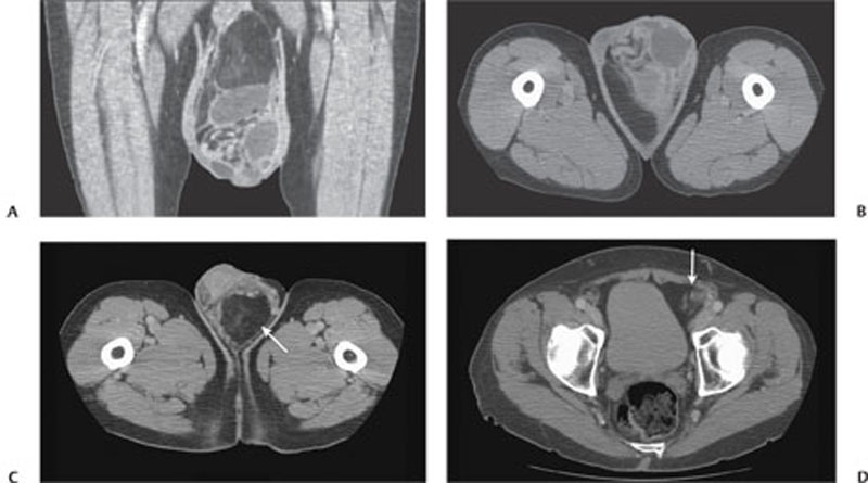
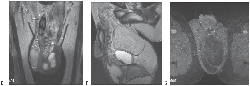
Radiologic Findings
Diagnosis
Related posts:


Stay updated, free articles. Join our Telegram channel

Full access? Get Clinical Tree


