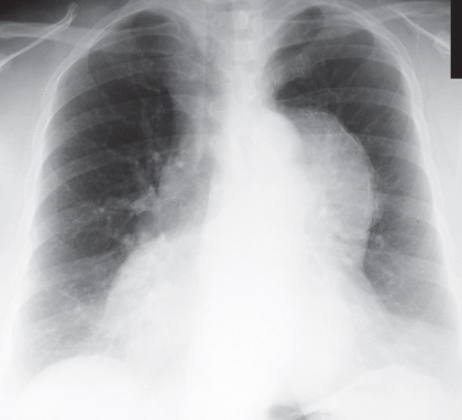
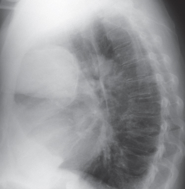
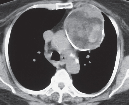
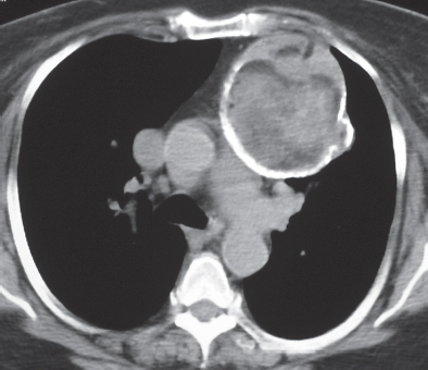
CASE 157 74-year-old woman with cough and chest pain PA (Fig. 157.1) and lateral (Fig. 157.2) chest radiographs demonstrate a large left anterior mediastinal mass with a thin peripheral rim of curvilinear calcification. Unenhanced chest CT (mediastinal window) (Figs. 157.3, 157.4) shows a heterogeneous spherical mass in the left lobe of the thymus. Note the peripheral curvilinear calcification and internal areas of soft tissue, fluid, and fat attenuation. Mature Teratoma None Germ cell neoplasms typically occur in the gonad but may also affect extragonadal sites, usually the anterior mediastinum (near or within the thymus). Mature teratoma is a benign neoplasm that accounts for approximately 70% of all primary mediastinal germ cell neoplasms. Fig. 157.1 Fig. 157.2 Fig. 157.3 Fig. 157.4 The etiology of germ cell neoplasms is poorly understood. It is theorized that extragonadal mature teratomas may arise from primitive germ cells that are “misplaced” along midline structures during their embryologic migration from the yolk sac to the gonad. Patients with mature teratoma are typically children and young adults (usually under 40 years of age). Patients may be asymptomatic or may present with signs and symptoms of compression of adjacent structures, including chest pain, dyspnea, and cough, or because of symptoms related to tumor rupture.
 Clinical Presentation
 Radiologic Findings
 Diagnosis
 Differential Diagnosis
 Discussion
Background
Etiology
Clinical Findings
Imaging Findings
Chest Radiography
Related posts:
![]()
Stay updated, free articles. Join our Telegram channel

Full access? Get Clinical Tree


Radiology Key
Fastest Radiology Insight Engine


